Explore
 Validate
 Learn
Learn Western blot
Antibody data
- Antibody Data
- Antigen structure
- References [1]
- Comments [0]
- Validations
- Western blot [2]
- Immunocytochemistry [1]
Submit
Validation data
Reference
Comment
Report error
- Product number
- PA3-007 - Provider product page

- Provider
- Invitrogen Antibodies
- Product name
- Anti-PDIR Polyclonal Antibody
- Antibody type
- Polyclonal
- Antigen
- Synthetic peptide
- Description
- PA3-007 detects PDIR protein in human samples. PA3-007 has successfully been used in Western blot procedures. By Western blot, this antibody detects an ~58 kDa protein representing PDIR in human fibrosarcoma HT 1080 cell lysate. To a lesser extent, this antibody also detects two unknown, nonspecific bands at 47 and 30 kDa. The PA3-007 immunogen is a synthetic peptide corresponding to residues T(498) N Y I R A L R E G D H E R L G K K(515) of human PDIR. This peptide (Cat. # PEP-226) is available for use in neutralization and control experiments.
- Reactivity
- Human
- Host
- Rabbit
- Isotype
- IgG
- Vial size
- 100 µL
- Concentration
- Conc. Not Determined
- Storage
- -20° C, Avoid Freeze/Thaw Cycles
Submitted references ERp18 regulates activation of ATF6α during unfolded protein response.
Oka OB, van Lith M, Rudolf J, Tungkum W, Pringle MA, Bulleid NJ
The EMBO journal 2019 Aug 1;38(15):e100990
The EMBO journal 2019 Aug 1;38(15):e100990
No comments: Submit comment
Supportive validation
- Submitted by
- Invitrogen Antibodies (provider)
- Main image
- Experimental details
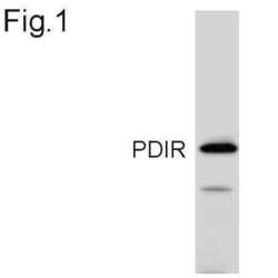
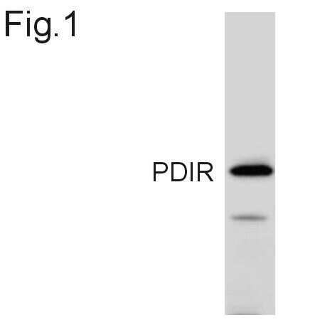
- Western blot of PDIR in HT 1080 cell lysate using Product # PA3-007.
- Submitted by
- Invitrogen Antibodies (provider)
- Main image

- Experimental details
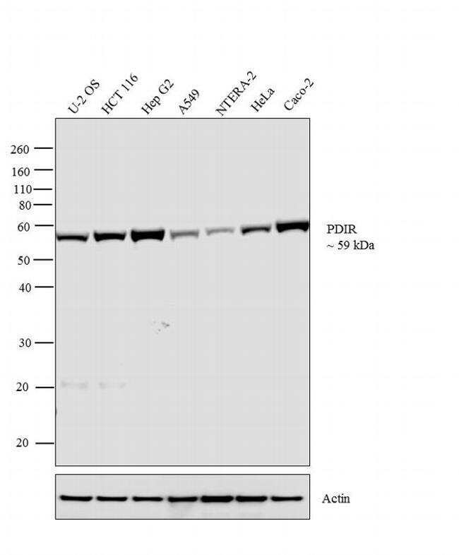
- Western blot analysis was performed on membrane enriched extracts (30 µglysate) of U-2 OS (Lane 1), HCT 116 (Lane 2), Hep G2 (Lane 3), A549 (Lane 4), NTERA-2 (Lane 5), HeLa (Lane 6) and Caco-2 (Lane 7). The blot was probed with Anti-PDIR Rabbit Polyclonal Antibody (Product # PA3-007, 1:1,000 dilution) and detected by chemiluminescence using Goat anti-Rabbit IgG (H+L) Superclonal™ Secondary Antibody, HRP conjugate (Product # A27036, 0.4 µg/mL, 1:2500 dilution). A 59 kDa band corresponding to PDIR was observed across the cell lines tested. Known quantity of protein samples were electrophoresed using Novex® NuPAGE® 10 % Bis-Tris gel (Product # NP0302BOX), XCell SureLock™ Electrophoresis System (Product # EI0002) and Novex® Sharp Pre-Stained Protein Standard (Product # LC5800). Resolved proteins were then transferred onto a nitrocellulose membrane with iBlot® 2 Dry Blotting System (Product # IB21001). The membrane was probed with the relevant primary and secondary Antibody following blocking with 5 % skimmed milk. Chemiluminescent detection was performed using Pierce™ ECL Western Blotting Substrate (Product # 32106).
Supportive validation
- Submitted by
- Invitrogen Antibodies (provider)
- Main image
- Experimental details
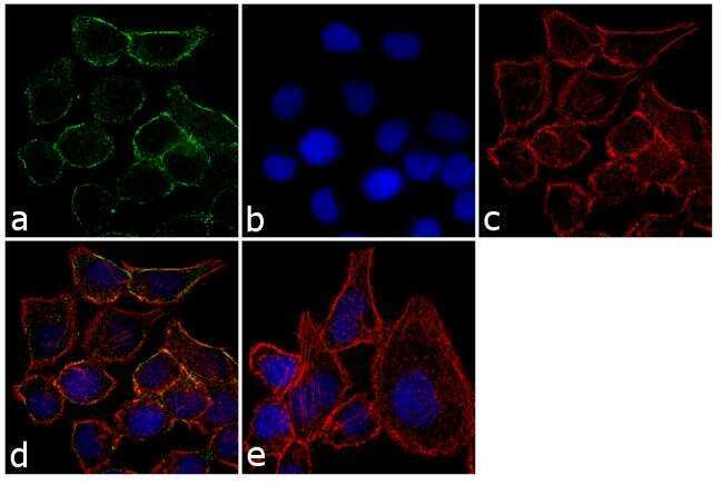
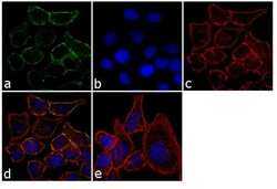
- Immunofluorescence analysis of PDIR was performed using 70% confluent log phase U-2 OS cells. The cells were fixed with 4% paraformaldehyde for 10 minutes, permeabilized with 0.1% Triton™ X-100 for 10 minutes, and blocked with 1% BSA for 1 hour at room temperature. The cells were labeled with PDIR Rabbit Polyclonal Antibody (Product # PA3-007) at 1:250 dilution in 0.1% BSA and incubated for 3 hours at room temperature and then labeled with Goat anti-Rabbit IgG (H+L) Superclonal™ Secondary Antibody, Alexa Fluor® 488 conjugate (Product # A27034) at a dilution of 1:2000 for 45 minutes at room temperature (Panel a: green). Nuclei (Panel b: blue) were stained with SlowFade® Gold Antifade Mountant with DAPI (Product # S36938). F-actin (Panel c: red) was stained with Rhodamine Phalloidin (Product # R415, 1:300). Panel d represents the merged image showing cytoplasmic localization. Panel e shows the no primary antibody control. The images were captured at 60X magnification.