Explore
 Validate
 Learn
Learn Western blot
 Immunocytochemistry
Antibody data
- Antibody Data
- Antigen structure
- References [0]
- Comments [0]
- Validations
- Western blot [4]
- Immunoprecipitation [1]
- Immunohistochemistry [2]
Submit
Validation data
Reference
Comment
Report error
- Product number
- NBP1-32213 - Provider product page

- Provider
- Novus Biologicals
- Proper citation
- Novus Cat#NBP1-32213, RRID:AB_10003918
- Product name
- Rabbit Polyclonal p27/Kip1 Antibody
- Antibody type
- Polyclonal
- Description
- Immunogen affinity purified.
- Reactivity
- Human, Mouse
- Host
- Rabbit
- Isotype
- IgG
- Vial size
- 0.1 ml
- Storage
- Aliquot and store at -20C or -80C. Avoid freeze-thaw cycles.
No comments: Submit comment
Supportive validation
- Submitted by
- Novus Biologicals (provider)
- Main image

- Experimental details
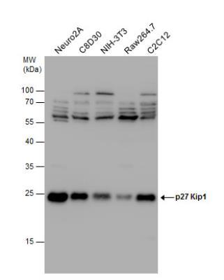
- Western Blot: p27/Kip1 Antibody [NBP1-32213] - Various whole cell extracts (30 ug) were separated by 12% SDS-PAGE, and the membrane was blotted with p27 Kip1 antibody diluted at 1:1000.
- Submitted by
- Novus Biologicals (provider)
- Main image
- Experimental details
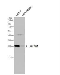
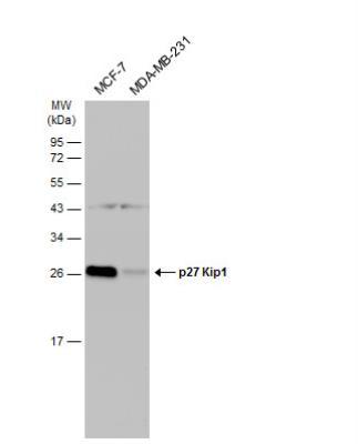
- Western Blot: p27/Kip1 Antibody [NBP1-32213] - Various whole cell extracts (30 ug) were separated by 12% SDS-PAGE, and the membrane was blotted with p27 Kip1 antibody diluted at 1:1000. The HRP-conjugated anti-rabbit IgG antibody (NBP2-19301) was used to detect the primary antibody.
- Submitted by
- Novus Biologicals (provider)
- Main image

- Experimental details
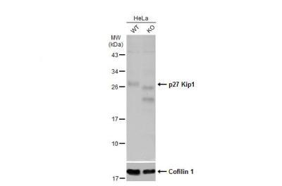
- Western Blot: p27/Kip1 Antibody [NBP1-32213] - Wild-type (WT) and p27 Kip1 knockout (KO) HeLa cell extracts (30 ug) were separated by 12% SDS-PAGE, and the membrane was blotted with p27 Kip1 antibody diluted at 1:500. HRP-conjugated anti-rabbit IgG antibody was used to detect the primary antibody.
- Submitted by
- Novus Biologicals (provider)
- Main image

- Experimental details
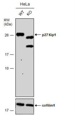
- Western Blot: p27/Kip1 Antibody [NBP1-32213] - Wild-type (WT) and p27 Kip1 knockout (KO) HeLa cell extracts (30 ug) were separated by 12% SDS-PAGE, and the membrane was blotted with p27 Kip1 antibody diluted at 1:500. HRP-conjugated anti-rabbit IgG antibody was used to detect the primary antibody.
Supportive validation
- Submitted by
- Novus Biologicals (provider)
- Main image

- Experimental details
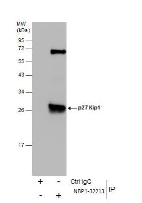
- Immunoprecipitation: p27/Kip1 Antibody [NBP1-32213] - Immunoprecipitation of p27 Kip1 protein from MCF-7 whole cell extracts using 5 ug of p27 Kip1 antibody. Western blot analysis was performed using p27 Kip1 antibody. EasyBlot anti-Rabbit IgG was used as a secondary reagent.
Supportive validation
- Submitted by
- Novus Biologicals (provider)
- Main image
- Experimental details
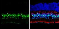
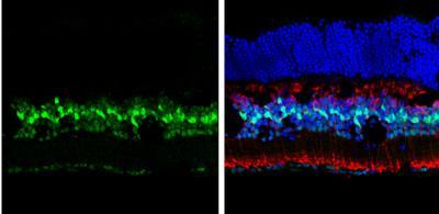
- Immunohistochemistry-Frozen: p27/Kip1 Antibody [NBP1-32213] - Frozen sectioned adult mouse retina. Green: p27 Kip1 protein stained by p27 Kip1 antibody diluted at 1:250. Red: Protein kinase C alpha staining. Blue: Fluoroshield with DAPI.
- Submitted by
- Novus Biologicals (provider)
- Main image

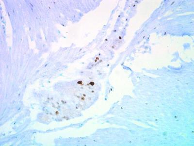
- Experimental details
- Immunohistochemistry-Paraffin: p27/Kip1 Antibody [NBP1-32213] - Staining of human ulcerative colitis tissue using anti-p27 Kip1 antibody.