 Explore
 Validate
 Learn
Learn Immunohistochemistry
Antibody data
- Antibody Data
- Antigen structure
- References [10]
- Comments [0]
- Validations
- Immunohistochemistry [3]
Submit
Validation data
Reference
Comment
Report error
- Product number
- PA5-16717 - Provider product page

- Provider
- Invitrogen Antibodies
- Product name
- p27 Kip1 Polyclonal Antibody
- Antibody type
- Polyclonal
- Antigen
- Synthetic peptide
- Description
- Based on 100% sequence identity, this antibody is predicted to react with Bovine and Cat
- Reactivity
- Human, Mouse
- Host
- Rabbit
- Isotype
- IgG
- Vial size
- 500 µL
- Storage
- Store at 4°C short term. For long term storage, store at -20°C, avoiding freeze/thaw cycles.
Submitted references Decreased blood vessel density and endothelial cell subset dynamics during ageing of the endocrine system.
In Vitro Models of GJB2-Related Hearing Loss Recapitulate Ca(2+) Transients via a Gap Junction Characteristic of Developing Cochlea.
The expression of cell cycle related proteins PCNA, Ki67, p27 and p57 in normal and preeclamptic human placentas.
Immunohistochemical distribution of cell cycle proteins p27, p57, cyclin D3, PCNA and Ki67 in normal and diabetic human placentas.
Immunolocalization of cell cycle proteins (p57, p27, cyclin D3, PCNA and Ki67) in intrauterine growth retardation (IUGR) and normal human term placentas.
Expression of anti-Müllerian hormone, CDKN1B, connexin 43, androgen receptor and steroidogenic enzymes in the equine cryptorchid testis.
Expression of insulin-like growth factor binding proteins during mouse cochlear development.
The diagnostic value of p27 in comparison to p57 in differentiation between different gestational trophoblastic diseases.
Expression of p27, COX-2, MLH1, and MSH2 in young patients with colon carcinoma and correlation with morphologic findings.
Immunolocalization of PCNA, Ki67, p27 and p57 in normal and dexamethasone-induced intrauterine growth restriction placental development in rat.
Chen J, Lippo L, Labella R, Tan SL, Marsden BD, Dustin ML, Ramasamy SK, Kusumbe AP
The EMBO journal 2021 Jan 4;40(1):e105242
The EMBO journal 2021 Jan 4;40(1):e105242
In Vitro Models of GJB2-Related Hearing Loss Recapitulate Ca(2+) Transients via a Gap Junction Characteristic of Developing Cochlea.
Fukunaga I, Fujimoto A, Hatakeyama K, Aoki T, Nishikawa A, Noda T, Minowa O, Kurebayashi N, Ikeda K, Kamiya K
Stem cell reports 2016 Dec 13;7(6):1023-1036
Stem cell reports 2016 Dec 13;7(6):1023-1036
The expression of cell cycle related proteins PCNA, Ki67, p27 and p57 in normal and preeclamptic human placentas.
Unek G, Ozmen A, Mendilcioglu I, Simsek M, Korgun ET
Tissue & cell 2014 Jun;46(3):198-205
Tissue & cell 2014 Jun;46(3):198-205
Immunohistochemical distribution of cell cycle proteins p27, p57, cyclin D3, PCNA and Ki67 in normal and diabetic human placentas.
Unek G, Ozmen A, Mendilcioglu I, Simsek M, Korgun ET
Journal of molecular histology 2014 Feb;45(1):21-34
Journal of molecular histology 2014 Feb;45(1):21-34
Immunolocalization of cell cycle proteins (p57, p27, cyclin D3, PCNA and Ki67) in intrauterine growth retardation (IUGR) and normal human term placentas.
Unek G, Ozmen A, Ozekinci M, Sakinci M, Korgun ET
Acta histochemica 2014 Apr;116(3):493-502
Acta histochemica 2014 Apr;116(3):493-502
Expression of anti-Müllerian hormone, CDKN1B, connexin 43, androgen receptor and steroidogenic enzymes in the equine cryptorchid testis.
Almeida J, Conley AJ, Ball BA
Equine veterinary journal 2013 Sep;45(5):538-45
Equine veterinary journal 2013 Sep;45(5):538-45
Expression of insulin-like growth factor binding proteins during mouse cochlear development.
Okano T, Kelley MW
Developmental dynamics : an official publication of the American Association of Anatomists 2013 Oct;242(10):1210-21
Developmental dynamics : an official publication of the American Association of Anatomists 2013 Oct;242(10):1210-21
The diagnostic value of p27 in comparison to p57 in differentiation between different gestational trophoblastic diseases.
Abdou A, Kandil M, El-Wahed MA, Shabaan M, El-Sharkawy M
Fetal and pediatric pathology 2013 Dec;32(6):395-411
Fetal and pediatric pathology 2013 Dec;32(6):395-411
Expression of p27, COX-2, MLH1, and MSH2 in young patients with colon carcinoma and correlation with morphologic findings.
Kenney B, Deng Y, Mitchell K
Human pathology 2013 Apr;44(4):591-7
Human pathology 2013 Apr;44(4):591-7
Immunolocalization of PCNA, Ki67, p27 and p57 in normal and dexamethasone-induced intrauterine growth restriction placental development in rat.
Unek G, Ozmen A, Kipmen-Korgun D, Korgun ET
Acta histochemica 2012 Jan;114(1):31-40
Acta histochemica 2012 Jan;114(1):31-40
No comments: Submit comment
Supportive validation
- Submitted by
- Invitrogen Antibodies (provider)
- Main image

- Experimental details
- Formalin-fixed, paraffin-embedded human prostate stained with p27 using peroxidase-conjugate and AEC chromogen. Note nuclear staining of glandular epithilium.

- Submitted by
- Invitrogen Antibodies (provider)
- Main image

- Experimental details
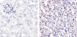
- Immunohistochemistry analysis of p27Kip1 showing staining in the nucleus of paraffin-embedded human prostate tissue (right) compared to a negative control without primary antibody (left). To expose target proteins, antigen retrieval was performed using 10mM sodium citrate (pH 6.0), microwaved for 8-15 min. Following antigen retrieval, tissues were blocked in 3% H2O2-methanol for 15 min at room temperature, washed with ddH2O and PBS, and then probed with a p27Kip1 Rabbit Polyclonal Antibody (Product # PA5-16717) diluted in 3% BSA-PBS at a dilution of 1:100 for 1 hour at 37°C in a humidified chamber. Tissues were washed extensively in PBST and detection was performed using an HRP-conjugated secondary antibody followed by colorimetric detection using a DAB kit. Tissues were counterstained with hematoxylin and dehydrated with ethanol and xylene to prep for mounting.

- Submitted by
- Invitrogen Antibodies (provider)
- Main image
- Experimental details
- Immunohistochemistry analysis of p27Kip1 showing staining in the nucleus of paraffin-embedded mouse kidney tissue (right) compared to a negative control without primary antibody (left). To expose target proteins, antigen retrieval was performed using 10mM sodium citrate (pH 6.0), microwaved for 8-15 min. Following antigen retrieval, tissues were blocked in 3% H2O2-methanol for 15 min at room temperature, washed with ddH2O and PBS, and then probed with a p27Kip1 Rabbit Polyclonal Antibody (Product # PA5-16717) diluted in 3% BSA-PBS at a dilution of 1:20 for 1 hour at 37°C in a humidified chamber. Tissues were washed extensively in PBST and detection was performed using an HRP-conjugated secondary antibody followed by colorimetric detection using a DAB kit. Tissues were counterstained with hematoxylin and dehydrated with ethanol and xylene to prep for mounting.
