Explore
 Validate
 Learn
Learn Western blot
 Immunohistochemistry
Antibody data
- Antibody Data
- Antigen structure
- References [1]
- Comments [0]
- Validations
- Western blot [1]
- Immunohistochemistry [1]
Submit
Validation data
Reference
Comment
Report error
- Product number
- HPA038719 - Provider product page

- Provider
- Atlas Antibodies
- Proper citation
- Atlas Antibodies Cat#HPA038719, RRID:AB_10670787
- Product name
- Anti-ACRV1
- Antibody type
- Polyclonal
- Description
- Polyclonal Antibody against Human ACRV1, Gene description: acrosomal vesicle protein 1, Alternative Gene Names: D11S4365, SP-10, SPACA2, Validated applications: IHC, WB, Uniprot ID: P26436, Storage: Store at +4°C for short term storage. Long time storage is recommended at -20°C.
- Reactivity
- Human
- Host
- Rabbit
- Conjugate
- Unconjugated
- Isotype
- IgG
- Vial size
- 100 µl
- Concentration
- 0.05 mg/ml
- Storage
- Store at +4°C for short term storage. Long time storage is recommended at -20°C.
- Handling
- The antibody solution should be gently mixed before use.
Submitted references The human testis-specific proteome defined by transcriptomics and antibody-based profiling
Djureinovic D, Fagerberg L, Hallström B, Danielsson A, Lindskog C, Uhlén M, Pontén F
MHR: Basic science of reproductive medicine 2014;20(6):476-488
MHR: Basic science of reproductive medicine 2014;20(6):476-488
No comments: Submit comment
Enhanced validation
- Submitted by
- Atlas Antibodies (provider)
- Enhanced method
- Recombinant expression validation
- Main image
- Experimental details
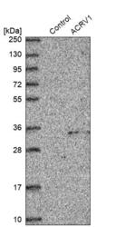
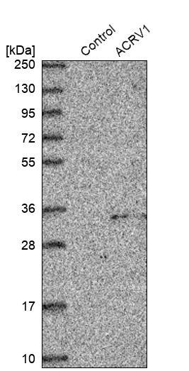
- Western blot analysis in control (vector only transfected HEK293T lysate) and ACRV1 over-expression lysate (Co-expressed with a C-terminal myc-DDK tag (~3.1 kDa) in mammalian HEK293T cells, LY412644).
- Sample type
- Human
- Protocol
- Protocol
Supportive validation
- Submitted by
- Atlas Antibodies (provider)
- Enhanced method
- Orthogonal validation
- Main image
- Experimental details
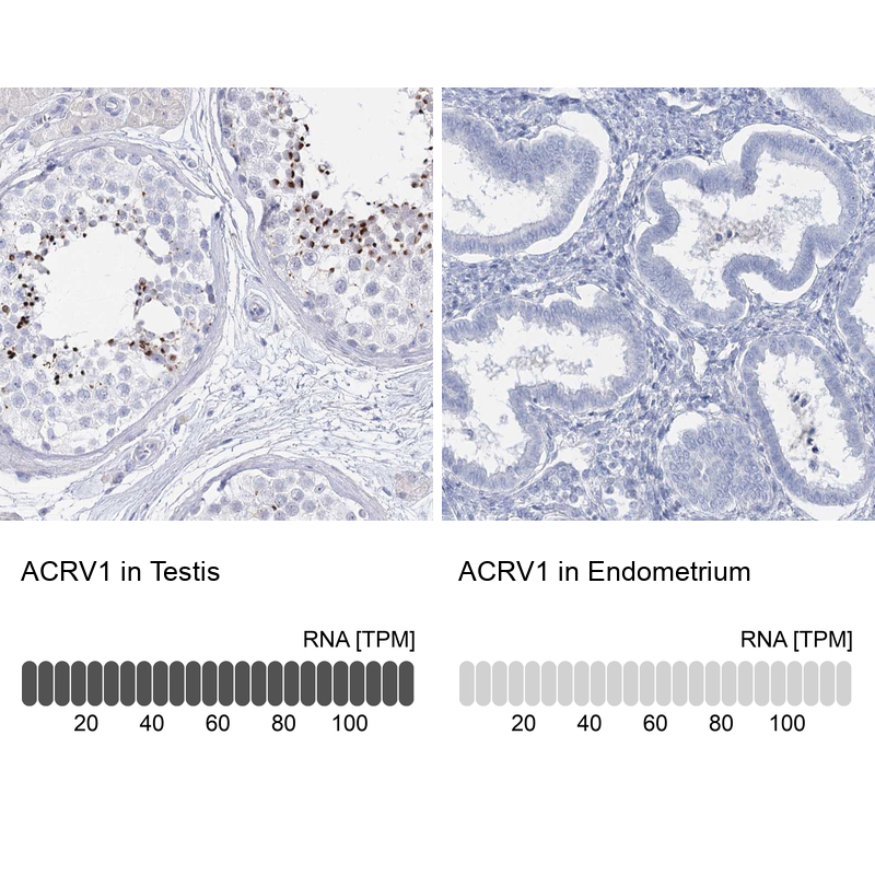
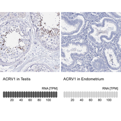
- Immunohistochemistry analysis in human testis and endometrium tissues using Anti-ACRV1 antibody. Corresponding ACRV1 RNA-seq data are presented for the same tissues.
- Sample type
- Human
- Protocol
- Protocol