Explore
 Validate
 Learn
LearnHPA029729
antibody from Atlas Antibodies
Targeting: HBS1L
DKFZp434g247, EF-1a, eRF3c, ERFS, HBS1, HSPC276, KIAA1038
 Western blot
 Immunocytochemistry
Antibody data
- Antibody Data
- Antigen structure
- References [3]
- Comments [0]
- Validations
- Western blot [1]
- Immunocytochemistry [1]
Submit
Validation data
Reference
Comment
Report error
- Product number
- HPA029729 - Provider product page

- Provider
- Atlas Antibodies
- Proper citation
- Atlas Antibodies Cat#HPA029729, RRID:AB_10601228
- Product name
- Anti-HBS1L
- Antibody type
- Polyclonal
- Description
- Polyclonal Antibody against Human HBS1L, Gene description: HBS1-like translational GTPase, Alternative Gene Names: DKFZp434g247, EF-1a, eRF3c, ERFS, HBS1, HSPC276, KIAA1038, Validated applications: WB, ICC, Uniprot ID: Q9Y450, Storage: Store at +4°C for short term storage. Long time storage is recommended at -20°C.
- Reactivity
- Human
- Host
- Rabbit
- Conjugate
- Unconjugated
- Isotype
- IgG
- Vial size
- 100 µl
- Concentration
- 0.1 mg/ml
- Storage
- Store at +4°C for short term storage. Long time storage is recommended at -20°C.
- Handling
- The antibody solution should be gently mixed before use.
Submitted references Landscape of functional interactions of human processive ribonucleases revealed by high-throughput siRNA screenings
A short splicing isoform of HBS1L links the cytoplasmic exosome and SKI complexes in humans
Systematic Analysis of Protein Pools, Isoforms, and Modifications Affecting Turnover and Subcellular Localization
Hojka-Osinska A, Chlebowski A, Grochowska J, Owczarek E, Affek K, Kłosowska-Kosicka K, Szczesny R, Dziembowski A
iScience 2021;24(9):103036
iScience 2021;24(9):103036
A short splicing isoform of HBS1L links the cytoplasmic exosome and SKI complexes in humans
Kalisiak K, Kuliński T, Tomecki R, Cysewski D, Pietras Z, Chlebowski A, Kowalska K, Dziembowski A
Nucleic Acids Research 2016
Nucleic Acids Research 2016
Systematic Analysis of Protein Pools, Isoforms, and Modifications Affecting Turnover and Subcellular Localization
Ahmad Y, Boisvert F, Lundberg E, Uhlen M, Lamond A
Molecular & Cellular Proteomics 2012;11(3):M111.013680
Molecular & Cellular Proteomics 2012;11(3):M111.013680
No comments: Submit comment
Enhanced validation
- Submitted by
- Atlas Antibodies (provider)
- Enhanced method
- Genetic validation
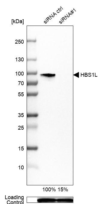
- Main image

- Experimental details
- Western blot analysis in U-251MG cells transfected with control siRNA, target specific siRNA probe #1, using Anti-HBS1L antibody. Remaining relative intensity is presented. Loading control: Anti-GAPDH.
- Sample type
- Human
- Protocol
- Protocol
Supportive validation
- Submitted by
- Atlas Antibodies (provider)
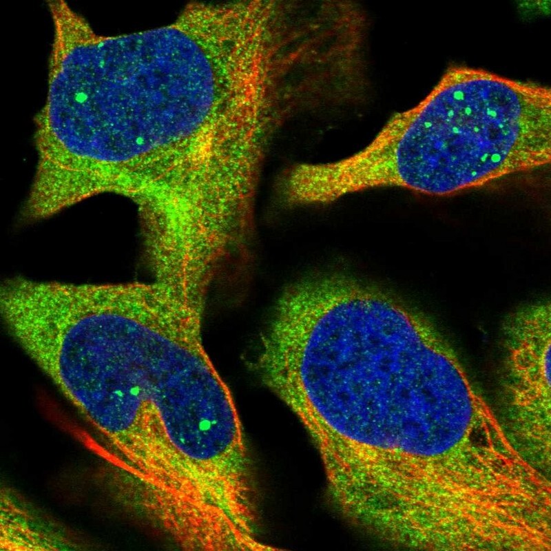
- Main image

- Experimental details
- Immunofluorescent staining of human cell line U-2 OS shows localization to nuclear bodies & cytosol.
- Sample type
- Human