Explore
 Validate
 Learn
Learn Western blot
Antibody data
- Antibody Data
- Antigen structure
- References [0]
- Comments [0]
- Validations
- Western blot [1]
- Immunocytochemistry [1]
- Flow cytometry [1]
Submit
Validation data
Reference
Comment
Report error
- Product number
- PA5-19305 - Provider product page

- Provider
- Invitrogen Antibodies
- Product name
- Anti-POU3F3/BRN1/OCT8
- Antibody type
- Polyclonal
- Antigen
- Synthetic peptide sequence (HMLSHAHQWVTAL) corresponding to the internal amino acids of POU3F3
- Reactivity
- Rat
- Host
- Goat
- Vial size
- 100 ug
- Concentration
- 0.5 mg/ml
- Storage
- -20° C, Avoid Freeze/Thaw Cycles
No comments: Submit comment
Supportive validation
- Submitted by
- Invitrogen Antibodies (provider)
- Main image

- Experimental details
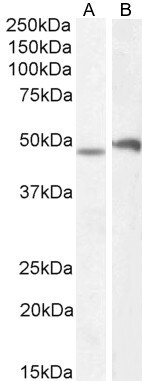
- Western blot analysis of POU3F3 using POU3F3 Polyclonal Antibody (Product # PA5-19305) (1 µg/mL) in staining of Mouse Spinal Cord (A) and Mouse Brain (B) lysate (35 µg protein in RIPA buffer). Detected by chemiluminescence.
Supportive validation
- Submitted by
- Invitrogen Antibodies (provider)
- Main image
- Experimental details
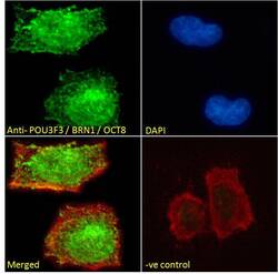
- Immunocytochemistry analysis of POU3F3 using POU3F3 Polyclonal Antibody (Product # PA5-19305) in paraformaldehyde fixed Neuro-2a cells, permeabilized with 0.15% Triton. Primary incubation 1hr (10 µg/mL) followed by Alexa Fluor 488 secondary antibody (2 µg/mL), showing nuclear staining. Actin filaments were stained with phalloidin (red) and the nuclear stain is DAPI (blue). Negative control: Unimmunized goat IgG (10 µg/mL) followed by Alexa Fluor 488 secondary antibody (2 µg/mL).
Supportive validation
- Submitted by
- Invitrogen Antibodies (provider)
- Main image
- Experimental details
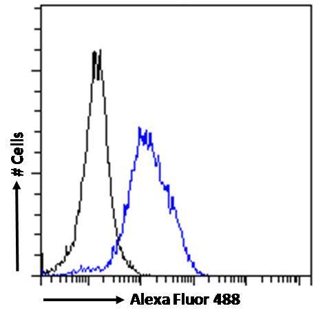
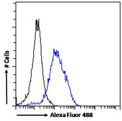
- Flow Cytometry analysis of POU3F3 using POU3F3 Polyclonal Antibody (Product # PA5-19305) in paraformaldehyde fixed Neuro-2a cells (blue line), permeabilized with 0.5% Triton. Primary incubation 1hr (10 µg/mL) followed by Alexa Fluor 488 secondary antibody (1 µg/mL). IgG control: Unimmunized goat IgG (black line) followed by Alexa Fluor 488 secondary antibody.