Explore
 Validate
 Learn
Learn Western blot
Antibody data
- Antibody Data
- Antigen structure
- References [0]
- Comments [0]
- Validations
- Western blot [2]
- Immunocytochemistry [1]
- Immunohistochemistry [5]
Submit
Validation data
Reference
Comment
Report error
- Product number
- HPA031123 - Provider product page

- Provider
- Atlas Antibodies
- Proper citation
- Atlas Antibodies Cat#HPA031123, RRID:AB_2301872
- Product name
- Anti-SLC12A8
- Antibody type
- Polyclonal
- Description
- Affinity purified using the PrEST antigen as affinity ligand
- Reactivity
- Human
- Host
- Rabbit
- Conjugate
- Unconjugated
- Antigen sequence
LLLEKAPSYGSEGPAQRVLEGTLLEFTKDMDQLLQ
LTRKLESSQPRQGEGNRTPESQKRKSKKATKQTLQ
DSFLLDLKSPPSFPVEISDRLPAASWEGQESCWNK
QTSKSEGTQPEGTYGEQLVPELCNQSESSGEDFFL
KSRLQ- Isotype
- IgG
- Vial size
- 100 µl
- Storage
- Store at +4°C for short term storage. Long time storage is recommended at -20°C.
No comments: Submit comment
Supportive validation
- Submitted by
- Atlas Antibodies (provider)
- Main image
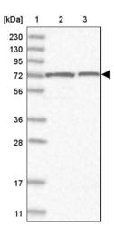
- Experimental details
- Lane 1: Marker [kDa] 230, 130, 95, 72, 56, 36, 28, 17, 11Lane 2: Human cell line RT-4Lane 3: Human cell line U-251MG sp

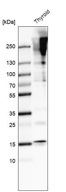
- Submitted by
- Atlas Antibodies (provider)
- Main image

- Experimental details
- Western blot analysis in human thyroid gland tissue.
- Sample type
- HUMAN
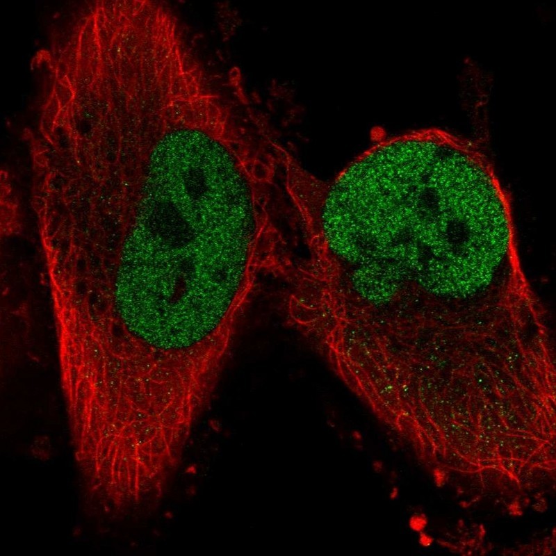
Supportive validation
- Submitted by
- Atlas Antibodies (provider)
- Main image

- Experimental details
- Immunofluorescent staining of human cell line U-251 MG shows localization to nucleoplasm.
- Sample type
- HUMAN
Supportive validation
- Submitted by
- Atlas Antibodies (provider)
- Main image
- Experimental details
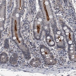
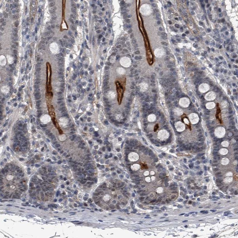
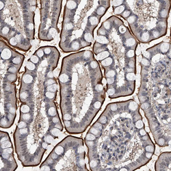
- Immunohistochemical staining of human duodenum shows strong positivity in luminal membrane.
- Submitted by
- Atlas Antibodies (provider)
- Main image

- Experimental details
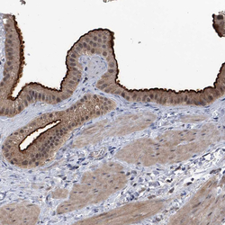
- Immunohistochemical staining of human prostate shows weak to moderate membranous positivity in glandular cells.
- Sample type
- HUMAN
- Submitted by
- Atlas Antibodies (provider)
- Main image
- Experimental details
- Immunohistochemical staining of human gallbladder shows strong membranous positivity in glandular cells.
- Sample type
- HUMAN

- Submitted by
- Atlas Antibodies (provider)
- Main image
- Experimental details
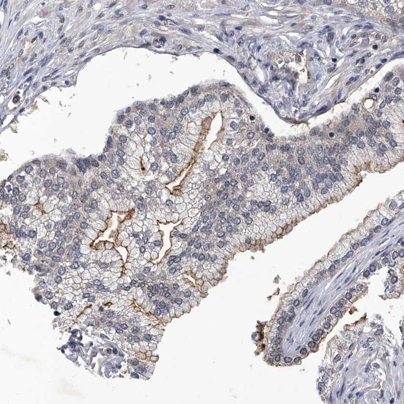
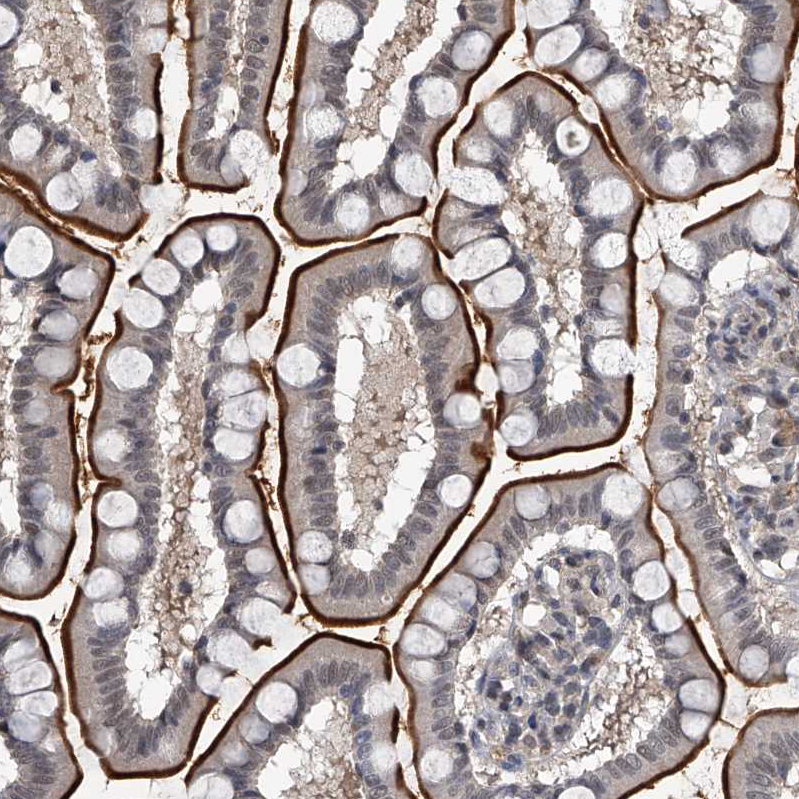
- Immunohistochemical staining of human upper gastrointestinal shows strong membranous positivity in glandular cells.
- Sample type
- HUMAN
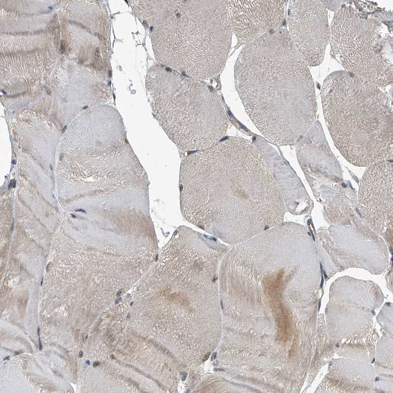
- Submitted by
- Atlas Antibodies (provider)
- Main image

- Experimental details
- Immunohistochemical staining of human skeletal muscle shows very weak to weak positivity in myocytes.
- Sample type
- HUMAN