Explore
 Validate
 Learn
Learn Western blot
 Immunocytochemistry
Antibody data
- Antibody Data
- Antigen structure
- References [0]
- Comments [0]
- Validations
- Western blot [1]
- Immunohistochemistry [1]
- Flow cytometry [1]
Submit
Validation data
Reference
Comment
Report error
- Product number
- NBP2-53421 - Provider product page

- Provider
- Novus Biologicals
- Product name
- Rabbit Polyclonal Lactate Dehydrogenase B Antibody
- Antibody type
- Polyclonal
- Description
- Immunogen affinity purified.
- Reactivity
- Human, Mouse, Rat
- Host
- Rabbit
- Isotype
- IgG
- Vial size
- 0.1 mg
- Concentration
- 1.0 mg/ml
- Storage
- Store at 4C short term. Aliquot and store at -20C long term. Avoid freeze-thaw cycles.
No comments: Submit comment
Supportive validation
- Submitted by
- Novus Biologicals (provider)
- Main image

- Experimental details
- Western Blot: Lactate Dehydrogenase B Antibody [NBP2-53421] - Total protein from human HeLa and A431 cells, mouse MEF cells and rat PC12 cells was separated on a 12% gel by SDS-PAGE, transferred to PVDF membrane and blocked in 5% non-fat milk in TBST. The membrane was probed with 1.0 ug/ml anti-LDHB in 1% non-fat milk in TBST and detected with an anti-rabbit HRP secondary antibody using chemiluminescence.

Supportive validation
- Submitted by
- Novus Biologicals (provider)
- Main image

- Experimental details
- Immunohistochemistry-Paraffin: Lactate Dehydrogenase B Antibody [NBP2-53421] - Analysis of a FFPE tissue section of mouse liver using Lactate Dehydrogenase B antibody at 1:400 dilution. The staining was developed using HRP-DAB detection method and the nuclei were counterstained with hematoxylin. The antibody generated a very specific staining in the hepatocytes.

Supportive validation
- Submitted by
- Novus Biologicals (provider)
- Main image
- Experimental details
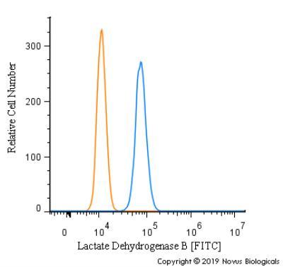
- Flow Cytometry: Lactate Dehydrogenase B Antibody [NBP2-53421] - An intracellular stain was performed on SK-MEL-28 cells with Lactate Dehyrdrogenase B Antibody NBP2-53421F (blue) and a matched isotype control (orange). Cells were fixed with 4% PFA and then permeabilized with 0.1% saponin. Cells were incubated in an antibody dilution of 5 ug/mL for 30 minutes at room temperature. Both antibodies were conjugated to FITC.