Explore
 Validate
 Learn
Learn Western blot
 Immunocytochemistry
Antibody data
- Antibody Data
- Antigen structure
- References [5]
- Comments [0]
- Validations
- Immunocytochemistry [1]
- Immunohistochemistry [2]
- Other assay [3]
Submit
Validation data
Reference
Comment
Report error
- Product number
- PA5-27505 - Provider product page

- Provider
- Invitrogen Antibodies
- Product name
- LDHB Polyclonal Antibody
- Antibody type
- Polyclonal
- Antigen
- Recombinant full-length protein
- Description
- Recommended positive controls: 293T, U87-MG, SK-N-SH, IMR32, SK-N-AS, mouse brain, rat brain. Predicted reactivity: Mouse (98%), Rat (98%), Xenopus laevis (81%), Dog (100%), Pig (96%), Rabbit (100%), Chicken (87%), Rhesus Monkey (100%), Chimpanzee (100%), Bovine (98%). Store product as a concentrated solution. Centrifuge briefly prior to opening the vial.
- Reactivity
- Human, Mouse, Rat
- Host
- Rabbit
- Isotype
- IgG
- Vial size
- 100 μL
- Concentration
- 0.32 mg/mL
- Storage
- Store at 4°C short term. For long term storage, store at -20°C, avoiding freeze/thaw cycles.
Submitted references LDHB Deficiency Promotes Mitochondrial Dysfunction Mediated Oxidative Stress and Neurodegeneration in Adult Mouse Brain.
R-2-hydroxyglutarate attenuates aerobic glycolysis in leukemia by targeting the FTO/m(6)A/PFKP/LDHB axis.
Quantitative Proteomic Approach Reveals Altered Metabolic Pathways in Response to the Inhibition of Lysine Deacetylases in A549 Cells under Normoxia and Hypoxia.
Branched-chain ketoacids secreted by glioblastoma cells via MCT1 modulate macrophage phenotype.
Lactate metabolism is associated with mammalian mitochondria.
Park JS, Saeed K, Jo MH, Kim MW, Lee HJ, Park CB, Lee G, Kim MO
Antioxidants (Basel, Switzerland) 2022 Jan 28;11(2)
Antioxidants (Basel, Switzerland) 2022 Jan 28;11(2)
R-2-hydroxyglutarate attenuates aerobic glycolysis in leukemia by targeting the FTO/m(6)A/PFKP/LDHB axis.
Qing Y, Dong L, Gao L, Li C, Li Y, Han L, Prince E, Tan B, Deng X, Wetzel C, Shen C, Gao M, Chen Z, Li W, Zhang B, Braas D, Ten Hoeve J, Sanchez GJ, Chen H, Chan LN, Chen CW, Ann D, Jiang L, Müschen M, Marcucci G, Plas DR, Li Z, Su R, Chen J
Molecular cell 2021 Mar 4;81(5):922-939.e9
Molecular cell 2021 Mar 4;81(5):922-939.e9
Quantitative Proteomic Approach Reveals Altered Metabolic Pathways in Response to the Inhibition of Lysine Deacetylases in A549 Cells under Normoxia and Hypoxia.
Martín-Bernabé A, Tarragó-Celada J, Cunin V, Michelland S, Cortés R, Poignant J, Boyault C, Rachidi W, Bourgoin-Voillard S, Cascante M, Seve M
International journal of molecular sciences 2021 Mar 25;22(7)
International journal of molecular sciences 2021 Mar 25;22(7)
Branched-chain ketoacids secreted by glioblastoma cells via MCT1 modulate macrophage phenotype.
Silva LS, Poschet G, Nonnenmacher Y, Becker HM, Sapcariu S, Gaupel AC, Schlotter M, Wu Y, Kneisel N, Seiffert M, Hell R, Hiller K, Lichter P, Radlwimmer B
EMBO reports 2017 Dec;18(12):2172-2185
EMBO reports 2017 Dec;18(12):2172-2185
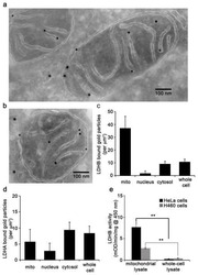
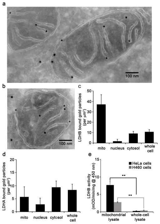
Lactate metabolism is associated with mammalian mitochondria.
Chen YJ, Mahieu NG, Huang X, Singh M, Crawford PA, Johnson SL, Gross RW, Schaefer J, Patti GJ
Nature chemical biology 2016 Nov;12(11):937-943
Nature chemical biology 2016 Nov;12(11):937-943
No comments: Submit comment
Supportive validation
- Submitted by
- Invitrogen Antibodies (provider)
- Main image
- Experimental details
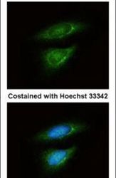
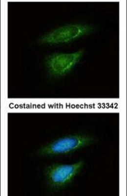
- Immunofluorescent analysis of LDH-B in methanol-fixed HeLa cells using a LDH-B polyclonal antibody (Product # PA5-27505) at a 1:50 dilution.
Supportive validation
- Submitted by
- Invitrogen Antibodies (provider)
- Main image

- Experimental details
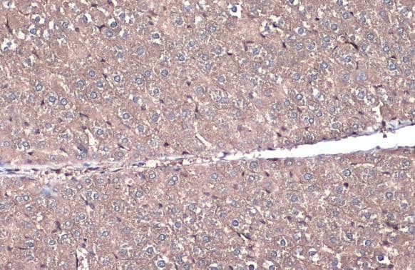
- LDHB Polyclonal Antibody detects LDH-B protein at cytoplasm by immunohistochemical analysis. Sample: Paraffin-embedded mouse liver. LDH-B stained by LDHB Polyclonal Antibody (Product # PA5-27505) diluted at 1:500. Antigen Retrieval: Citrate buffer, pH 6.0, 15 min.
- Submitted by
- Invitrogen Antibodies (provider)
- Main image

- Experimental details
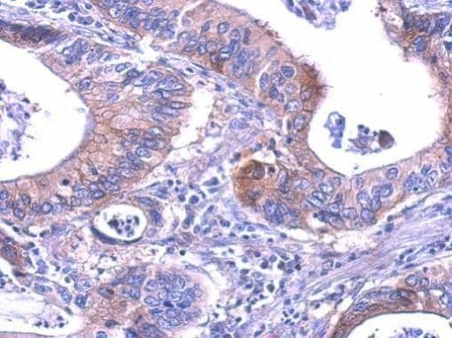
- LDHB Polyclonal Antibody detects LDHB protein at cytosol on human gastric cancer by immunohistochemical analysis. Sample: Paraffin-embedded gastric cancer. LDHB Polyclonal Antibody (Product # PA5-27505) dilution: 1:200. Antigen Retrieval: EDTA based buffer, pH 8.0, 15 min.
Supportive validation
- Submitted by
- Invitrogen Antibodies (provider)
- Main image
- Experimental details
- NULL
- Submitted by
- Invitrogen Antibodies (provider)
- Main image
- Experimental details
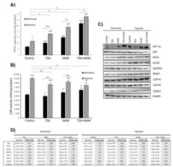
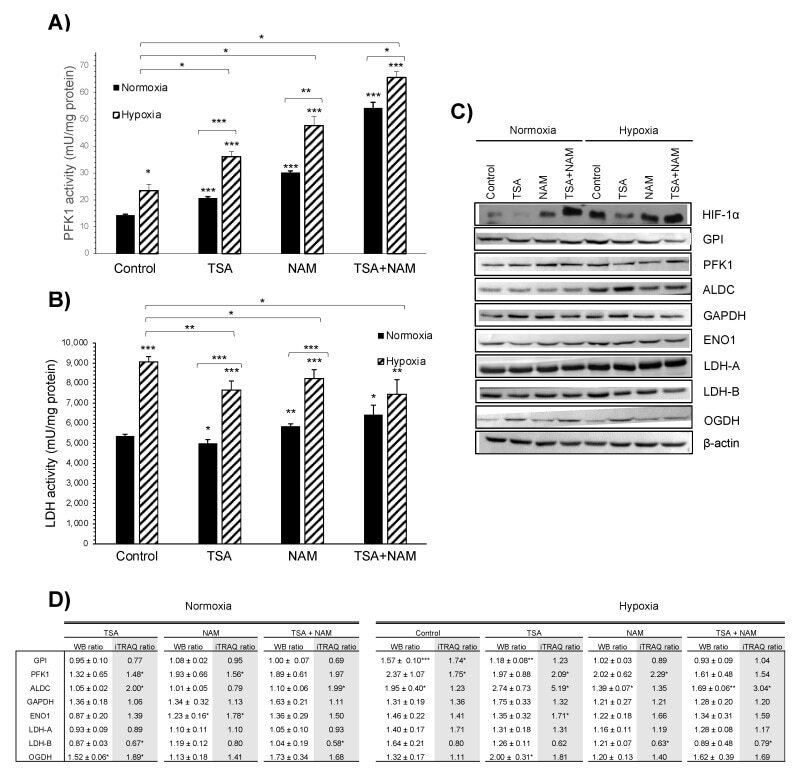
- Figure 4 Effect of KDAC inhibition on enzyme activities in A549 cells under normoxia and hypoxia. ( A , B ) The ATP- dependent 6-phosphofructokinase (PFK1) ( A ) and lactate dehydrogenase (LDH) ( B ) enzymatic activities were measured after 24 h of incubation, and activities were normalized to intracellular protein content in each condition. A549 cells were treated with 1 muM of TSA, 20 mM of NAM, and both 1 muM TSA and 20 mM NAM for 24 h of incubation under normoxia and hypoxia. Cells incubated in medium without KDACIs served as control. Bars represent the means +- standard error of the mean of three independent experiments. The asterisks above bars indicate statistically significant differences compared to normoxic control cells. The asterisks above curly brackets indicate statistically significant differences between hypoxic and normoxic treatments and between hypoxic treatments and hypoxic control cells. Statistical significance was assessed by a two-tailed Student''s t -test. *, p
- Submitted by
- Invitrogen Antibodies (provider)
- Main image
- Experimental details
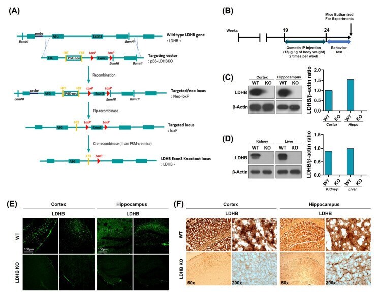
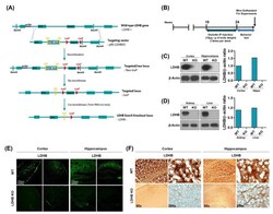
- Figure 1 Schematic diagram showing the generation of Ldhb -/- mouse, experimental design, and verification of Ldhb -/- model. ( A ) Schematic diagram illustrating the Cre-loxP and Flp-FRT system using pBS- Ldhb KO vectors. Ldhb -/- mice were generated through gene knockout at the exon 3 loci. ( B ) Schematic diagram of experimental design, showing the period of osmotin treatment in Ldhb -/- mice and behavior test. ( C , D ) The Western blot analysis showing the expression of LDHB in the cortex, hippocampus, kidney, and liver of Ldhb -/- and WT mice. beta-actin was used as a loading control. The bands were quantified using ImageJ software, and the differences are represented by histograms. ( E ) Confocal microscopic images showing the expression of LDHB in the cortex and hippocampus of Ldhb -/- , WT mice. ( F ) Immunohistochemistry staining for LDHB in the cortex and hippocampus of Ldhb -/- , WT mice.