Explore
 Validate
 Learn
Learn Western blot
Antibody data
- Antibody Data
- Antigen structure
- References [3]
- Comments [0]
- Validations
- Western blot [2]
- Immunohistochemistry [1]
Submit
Validation data
Reference
Comment
Report error
- Product number
- NB110-74570 - Provider product page

- Provider
- Novus Biologicals
- Proper citation
- Novus Cat#NB110-74570, RRID:AB_1050137
- Product name
- Mouse Monoclonal CHT1 Antibody
- Antibody type
- Monoclonal
- Description
- Tissue culture supernatant.
- Reactivity
- Human, Mouse, Rat, Simian
- Host
- Mouse
- Isotype
- IgG
- Vial size
- 0.1 ml
- Storage
- Store at 4C short term. Aliquot and store at -20C long term. Avoid freeze-thaw cycles.
Submitted references Aberrant trafficking of the high-affinity choline transporter in AP-3-deficient mice.
Vesicular localization and activity-dependent trafficking of presynaptic choline transporters.
Distribution of high affinity choline transporter immunoreactivity in the primate central nervous system.
Misawa H, Fujigaya H, Nishimura T, Moriwaki Y, Okuda T, Kawashima K, Nakata K, Ruggiero AM, Blakely RD, Nakatsu F, Ohno H
The European journal of neuroscience 2008 Jun;27(12):3109-17
The European journal of neuroscience 2008 Jun;27(12):3109-17
Vesicular localization and activity-dependent trafficking of presynaptic choline transporters.
Ferguson SM, Savchenko V, Apparsundaram S, Zwick M, Wright J, Heilman CJ, Yi H, Levey AI, Blakely RD
The Journal of neuroscience : the official journal of the Society for Neuroscience 2003 Oct 29;23(30):9697-709
The Journal of neuroscience : the official journal of the Society for Neuroscience 2003 Oct 29;23(30):9697-709
Distribution of high affinity choline transporter immunoreactivity in the primate central nervous system.
Kus L, Borys E, Ping Chu Y, Ferguson SM, Blakely RD, Emborg ME, Kordower JH, Levey AI, Mufson EJ
The Journal of comparative neurology 2003 Aug 25;463(3):341-57
The Journal of comparative neurology 2003 Aug 25;463(3):341-57
No comments: Submit comment
Supportive validation
- Submitted by
- Novus Biologicals (provider)
- Main image

- Experimental details
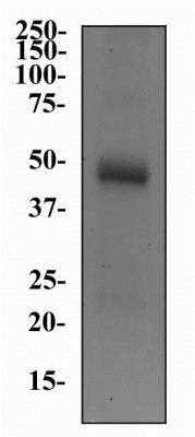
- Western Blot: CHT1 Antibody (62-2E8) [NB110-74570] - Western Blot Image of anti-CHT1 (62-2E8). Total protein from mouse brain was separated on a 12% gel by SDS-PAGE, transferred to PVDF membrane and blocked in 5% non-fat milk in TBST. The membrane was probed with a 1:500 dilution of anti-CHT1 in 1% milk/TBST. Reactivity was detected with an anti-mouse HRP secondary antibody using chemiluminescence.
- Submitted by
- Novus Biologicals (provider)
- Main image
- Experimental details
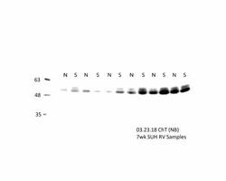
- Western Blot: CHT1 Antibody (62-2E8) [NB110-74570] - ChT blot with Rat RV tissue. N: Normoxia, S: SUH/SUGEN+Hypoxia. Total protein from rat heart (RV) was separated on a 10% gel by SDS-PAGE, transferred to PVDF membrane and blocked in 5% non-fat milk in TBS 0.1% Tween. The membrane was probed with a 1:500 dilution of anti-CHT1 in 5% milk/TBST overnight at 4C. Reactivity was detected with an anti-mouse HRP secondary antibody (1:5000) using chemiluminescence on Biorad imager. This image was submitted via customer review.

Supportive validation
- Submitted by
- Novus Biologicals (provider)
- Main image

- Experimental details
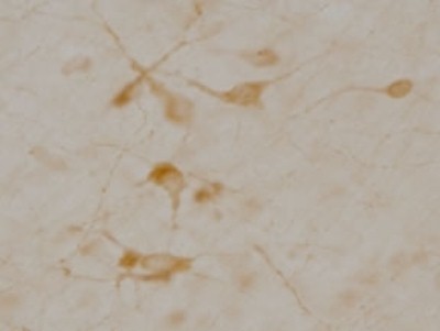
- Immunohistochemistry: CHT1 Antibody (62-2E8) [NB110-74570] - Staining of neurons in rat basal forebrain