Explore
 Validate
 Learn
Learn Western blot
Antibody data
- Antibody Data
- Antigen structure
- References [1]
- Comments [0]
- Validations
- Western blot [2]
- Immunohistochemistry [7]
- Flow cytometry [1]
Submit
Validation data
Reference
Comment
Report error
- Product number
- TA503467 - Provider product page

- Provider
- OriGene
- Proper citation
- OriGene Cat#TA503467, RRID:AB_11126826
- Product name
- HSPBP1 mouse monoclonal antibody, clone OTI1D5 (formerly 1D5)
- Antibody type
- Monoclonal
- Description
- HSPBP1 mouse monoclonal antibody, clone OTI1D5 (formerly 1D5)
- Host
- Mouse
- Conjugate
- Unconjugated
- Epitope
- HSPBP1
- Isotype
- IgG
- Antibody clone number
- OTI1D5
- Vial size
- 100 µl
- Concentration
- 0.43 mg/ml
Submitted references Sorcin induces gastric cancer cell migration and invasion contributing to STAT3 activation.
Tuo H, Shu F, She S, Yang M, Zou XQ, Huang J, Hu HD, Hu P, Ren H, Peng SF, Yang YX
Oncotarget 2017 Nov 28;8(61):104258-104271
Oncotarget 2017 Nov 28;8(61):104258-104271
No comments: Submit comment
Supportive validation
- Submitted by
- OriGene (provider)
- Main image
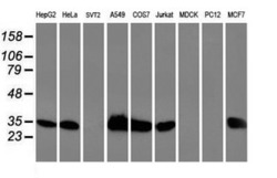
- Experimental details
- Western blot analysis of extracts (35ug) from 9 different cell lines by using anti-HSPBP1 monoclonal antibody.
- Validation comment
- WB

- Submitted by
- OriGene (provider)
- Main image
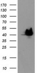
- Experimental details
- HEK293T cells were transfected with the pCMV6-ENTRY control (Left lane) or pCMV6-ENTRY HSPBP1 (RC201814, Right lane) cDNA for 48 hrs and lysed. Equivalent amounts of cell lysates (5 ug per lane) were separated by SDS-PAGE and immunoblotted with anti-HSPBP1.
- Validation comment
- WB

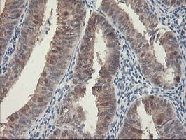
Supportive validation
- Submitted by
- OriGene (provider)
- Main image

- Experimental details
- Immunohistochemical staining of paraffin-embedded Adenocarcinoma of Human endometrium tissue using anti-HSPBP1 mouse monoclonal antibody. (Heat-induced epitope retrieval by 10mM citric buffer, pH6.0, 100C for 10min, TA503467)
- Validation comment
- IHC
- Submitted by
- OriGene (provider)
- Main image
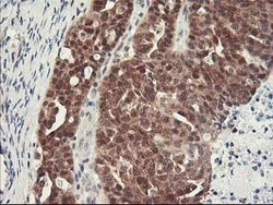
- Experimental details
- Immunohistochemical staining of paraffin-embedded Human pancreas tissue within the normal limits using anti-HSPBP1 mouse monoclonal antibody. (Heat-induced epitope retrieval by 10mM citric buffer, pH6.0, 100C for 10min, TA503467)
- Validation comment
- IHC

- Submitted by
- OriGene (provider)
- Main image
- Experimental details
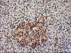
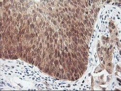
- Immunohistochemical staining of paraffin-embedded Adenocarcinoma of Human ovary tissue using anti-HSPBP1 mouse monoclonal antibody. (Heat-induced epitope retrieval by 10mM citric buffer, pH6.0, 100C for 10min, TA503467)
- Validation comment
- IHC

- Submitted by
- OriGene (provider)
- Main image
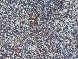
- Experimental details
- Immunohistochemical staining of paraffin-embedded Human lymphoma tissue using anti-HSPBP1 mouse monoclonal antibody. (Heat-induced epitope retrieval by 10mM citric buffer, pH6.0, 100C for 10min, TA503467)
- Validation comment
- IHC

- Submitted by
- OriGene (provider)
- Main image
- Experimental details
- Immunohistochemical staining of paraffin-embedded Human endometrium tissue within the normal limits using anti-HSPBP1 mouse monoclonal antibody. (Heat-induced epitope retrieval by 10mM citric buffer, pH6.0, 100C for 10min, TA503467)
- Validation comment
- IHC

- Submitted by
- OriGene (provider)
- Main image
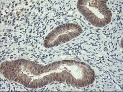
- Experimental details
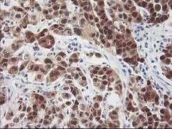
- Immunohistochemical staining of paraffin-embedded Carcinoma of Human bladder tissue using anti-HSPBP1 mouse monoclonal antibody. (Heat-induced epitope retrieval by 10mM citric buffer, pH6.0, 100C for 10min, TA503467)
- Validation comment
- IHC

- Submitted by
- OriGene (provider)
- Main image
- Experimental details
- Immunohistochemical staining of paraffin-embedded Carcinoma of Human lung tissue using anti-HSPBP1 mouse monoclonal antibody. (Heat-induced epitope retrieval by 10mM citric buffer, pH6.0, 100C for 10min, TA503467)
- Validation comment
- IHC

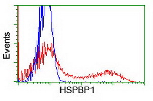
Supportive validation
- Submitted by
- OriGene (provider)
- Main image
- Experimental details
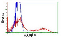
- HEK293T cells transfected with either RC201814 overexpress plasmid(Red) or empty vector control plasmid(Blue) were immunostained by anti-HSPBP1 antibody(TA503467), and then analyzed by flow cytometry.
- Validation comment
- FC