Explore
 Validate
 Learn
Learn Western blot
 Immunocytochemistry
 Immunohistochemistry
Antibody data
- Antibody Data
- Antigen structure
- References [1]
- Comments [0]
- Validations
- Immunocytochemistry [1]
- Other assay [3]
Submit
Validation data
Reference
Comment
Report error
- Product number
- MA5-36130 - Provider product page

- Provider
- Invitrogen Antibodies
- Product name
- ApoER2 Monoclonal Antibody (13B2)
- Antibody type
- Monoclonal
- Antigen
- Recombinant full-length protein
- Reactivity
- Human
- Host
- Mouse
- Isotype
- IgG
- Antibody clone number
- 13B2
- Vial size
- 100 μL
- Concentration
- 2 mg/mL
- Storage
- Store at 4°C short term. For long term storage, store at -20°C, avoiding freeze/thaw cycles.
Submitted references The apolipoprotein receptor LRP3 compromises APP levels.
Cuchillo-Ibañez I, Lennol MP, Escamilla S, Mata-Balaguer T, Valverde-Vozmediano L, Lopez-Font I, Ferrer I, Sáez-Valero J
Alzheimer's research & therapy 2021 Nov 2;13(1):181
Alzheimer's research & therapy 2021 Nov 2;13(1):181
No comments: Submit comment
Supportive validation
- Submitted by
- Invitrogen Antibodies (provider)
- Main image
- Experimental details
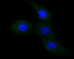
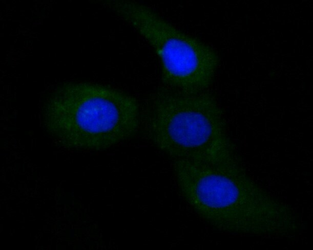
- Immunocytochemical analysis of ApoER2 in SKOV-3 cells using a ApoER2 monoclonal antibody (Product #MA5-36130). Formalin fixed cells were permeabilized with 0.1% Triton X-100 in TBS for 10 minutes at room temperature and blocked with 1% Blocker BSA for 15 minutes at room temperature. Cells were probed with the primary antibody for 1 hour at room temperature, washed with PBS. Alexa Fluor®488 Goat anti-Mouse IgG was used as the secondary antibody at 1:100 dilution. The nuclear counter stain is DAPI.
Supportive validation
- Submitted by
- Invitrogen Antibodies (provider)
- Main image
- Experimental details
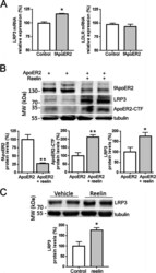
- Fig. 1 ApoER2/reelin signaling upregulates LRP3 expression. a qRT-PCR analysis showing expression of LRP3 mRNA and LDLR mRNA after transfection with GFP cDNA (control) and full-length ApoER2 cDNA (fApoER2) in SH-SYSH cells. 18S was used as an internal control for mRNA expression ( n = 10-12 for each condition, p < 0.001 for control versus fApoER2; t -test with Welch's correction). Note that the X axis begins at 50%. b Quantification and western blot showing the expression of full-length ApoER2, ApoER2-CTF, and LRP3 proteins after fApoER2 transfection and reelin (12 mug/ml) treatment for 24 h in SH-SY5Y cells. Tubulin was used as an internal control ( n = 9 for each condition, ** p < 0.001 for expression of fApoER2, t -test with Welch's correction, and ApoER2-CTF, t -test; * p < 0.05 for expression of LRP3, t -test). c Quantification and western blot showing the expression of LRP3 protein after reelin (12 mug/ml) treatment for 24 h or vehicle (Hanks's media) in neuro-differentiated SH-SY5Y cells with retinoic acid ( n = 9 for each condition, * p

- Submitted by
- Invitrogen Antibodies (provider)
- Main image
- Experimental details
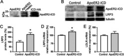
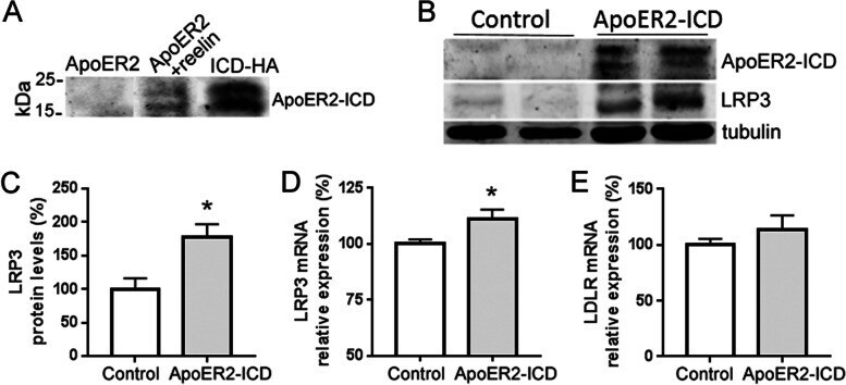
- Fig. 2 ApoER2-ICD increases LRP3 expression. a Representative western blot showing the expression of ApoER2-ICD after transfection with full-length ApoER2 cDNA (ApoER2) and reelin treatment (12mug/ml, ApoER2 + reelin) for 24 h in SH-SY5Y cells. For comparison, the expression of the ApoER2-ICD construct (ApoER2-ICD) is also shown. b Western blot and c quantification of LRP3 protein expression after ApoER2-ICD transfection in SH-SY5Y cells. Tubulin was used as an internal control ( n = 6 for each condition, * p < 0.001, t -test). d qRT-PCR analysis showing the expression of LRP3 mRNA ( n = 7 for each condition, * p < 0.05, t -test) and e LDLR mRNA ( n = 10 for each condition) after transfection with GFP cDNA (control) and ApoER2-ICD cDNA in SH-SY5Y cells. Note that the X axis in d begins at 50%. 18S was used as an internal control for mRNA expression
- Submitted by
- Invitrogen Antibodies (provider)
- Main image
- Experimental details
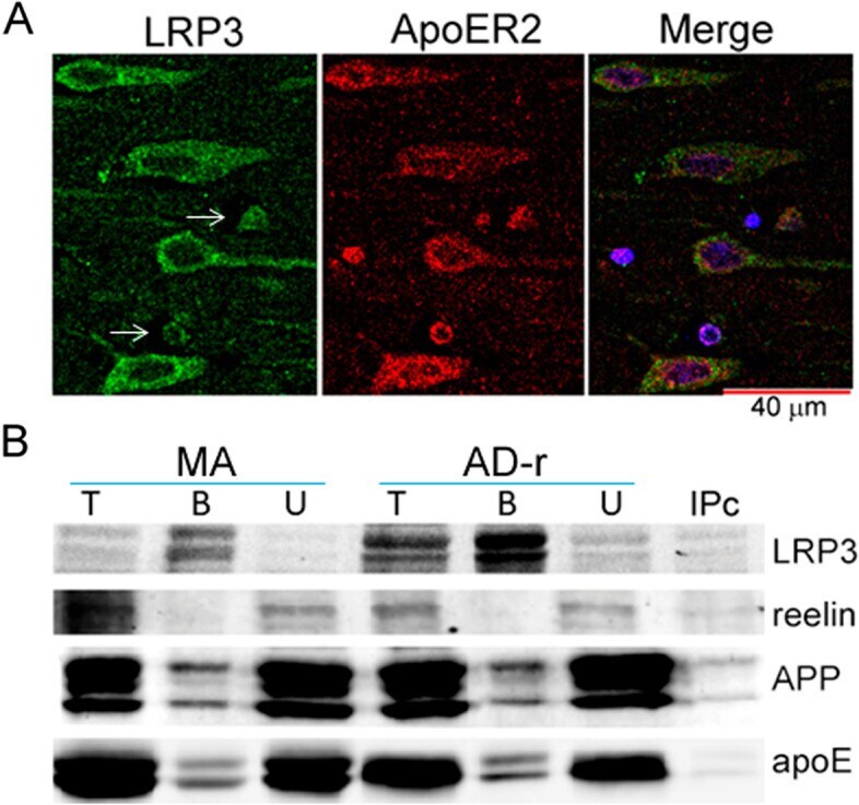
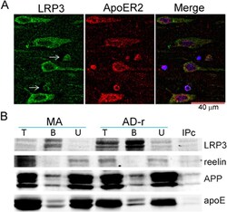
- Fig. 5 LRP3 co-immunoprecipitates with apoE and APP. a Representative immunofluorescence photomicrograph showing LRP3 and ApoER2 labeling in the same cells in a hippocampus slice of a MA subject. Neurons (large cells) show LRP3 and ApoER2 co-localization; in addition, oligodendroglia-like cells (thin arrows) also co-localize both antibodies. b Representative western blots showing immunoprecipitation of LRP3 and co-immunoprecipitation of reelin (no immunoprecipitation), apoE, and APP, from non-demented (ND) and Alzheimer's disease (AD) extracts. T total input, B bound fraction, U unbound fraction, IPc bound fraction of the negative control