Explore
 Validate
 Learn
Learn Immunohistochemistry
Antibody data
- Antibody Data
- Antigen structure
- References [2]
- Comments [0]
- Validations
- Immunohistochemistry [1]
- Other assay [4]
Submit
Validation data
Reference
Comment
Report error
- Product number
- PA5-64274 - Provider product page

- Provider
- Invitrogen Antibodies
- Product name
- ADAMTS8 Polyclonal Antibody
- Antibody type
- Polyclonal
- Antigen
- Recombinant full-length protein
- Description
- Immunogen sequence: SIATLERLQS FRPLPEPLTV QLLTVPGEVF PPKVKYTFFV PNDVDFSMQS SKERATTNII QPLLHAQWVL Highest antigen sequence identity to the following orthologs: Mouse - 83%, Rat - 83%.
- Reactivity
- Human
- Host
- Rabbit
- Isotype
- IgG
- Vial size
- 100 µL
- Concentration
- 0.05 mg/mL
- Storage
- Store at 4°C short term. For long term storage, store at -20°C, avoiding freeze/thaw cycles.
Submitted references METTL14 promotes tumorigenesis by regulating lncRNA OIP5-AS1/miR-98/ADAMTS8 signaling in papillary thyroid cancer.
Relationship Between ADAMTS8, ADAMTS18, and ADAMTS20 (A Disintegrin and Metalloproteinase with Thrombospondin Motifs) Expressions and Tumor Molecular Classification, Clinical Pathological Parameters, and Prognosis in Breast Invasive Ductal Carcinoma.
Zhang X, Li D, Jia C, Cai H, Lv Z, Wu B
Cell death & disease 2021 Jun 15;12(6):617
Cell death & disease 2021 Jun 15;12(6):617
Relationship Between ADAMTS8, ADAMTS18, and ADAMTS20 (A Disintegrin and Metalloproteinase with Thrombospondin Motifs) Expressions and Tumor Molecular Classification, Clinical Pathological Parameters, and Prognosis in Breast Invasive Ductal Carcinoma.
Guo X, Li J, Zhang H, Liu H, Liu Z, Wei X
Medical science monitor : international medical journal of experimental and clinical research 2018 Jun 3;24:3726-3735
Medical science monitor : international medical journal of experimental and clinical research 2018 Jun 3;24:3726-3735
No comments: Submit comment
Supportive validation
- Submitted by
- Invitrogen Antibodies (provider)
- Main image
- Experimental details
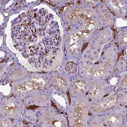
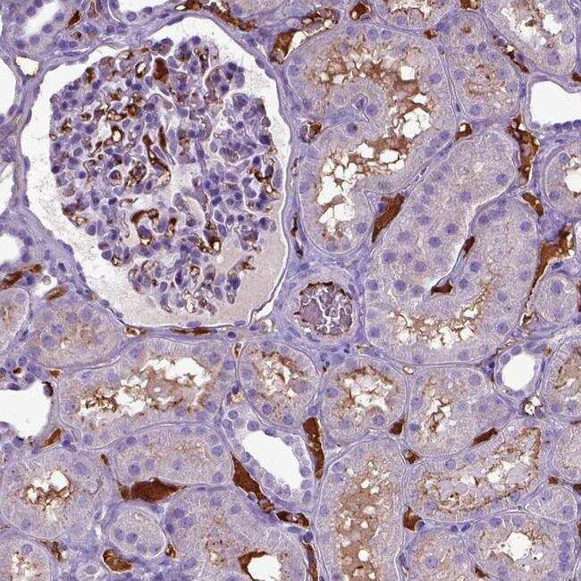
- Immunohistochemical staining of ADAMTS8 in human kidney tissue shows strong positivity in blood vessels and plasma. Samples were probed using an ADAMTS8 Polyclonal Antibody (Product # PA5-64274).
Supportive validation
- Submitted by
- Invitrogen Antibodies (provider)
- Main image
- Experimental details
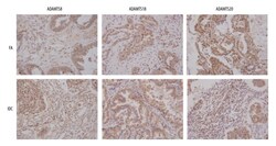
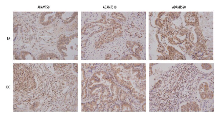
- Figure 1 IHC staining result of ADAMTS8, ADAMTS18, and ADAMTS20 in IDC and non-proliferative character of FA. In IDC, the positive expressions of ADAMTS8 and ADAMTS18 were significantly higher, while ADAMTS20 was lower, compared with those in FA. The sepia sections represent the stained proteins. The results showed that proteins were distributed in both cytoplasm and cytoplasm interstitial, mainly in cytoplasm.
- Submitted by
- Invitrogen Antibodies (provider)
- Main image

- Experimental details
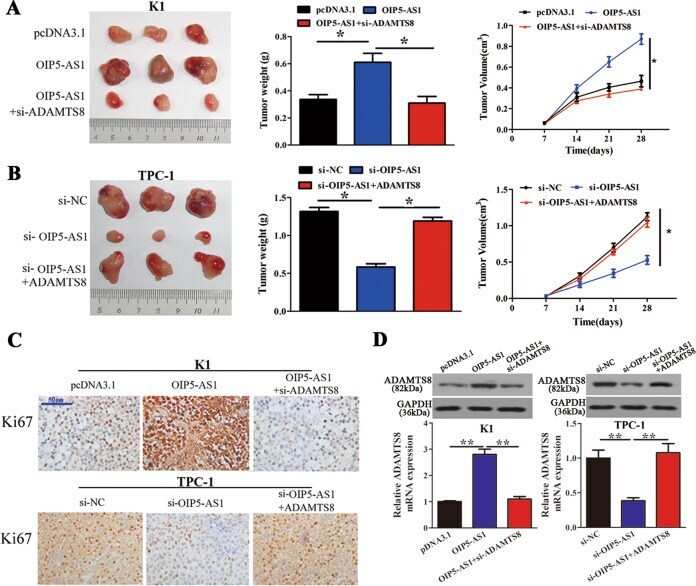
- Fig. 3 OIP5-AS1 regulates PTC cell growth in vivo. A Tumor weight and growth curves ( n = 6) were measured after injection of K1 cells transfected with OIP5-AS1 and si-ADAMTS8. B Tumor weight and growth curves ( n = 6) were measured after injection of TPC-1 cells transfected with si-OIP5-AS1 and ADAMTS8. C Ki67 staining to assess the proliferation capacity of K1 cells and TPC-1 cells ( n = 6). Scale bar = 50 mum. D Western blot and qRT-PCR analyses of ADAMTS8 expression levels in K1 and TPC-1 cells ( n = 6). * p < 0.05 and ** p < 0.01.
- Submitted by
- Invitrogen Antibodies (provider)
- Main image
- Experimental details
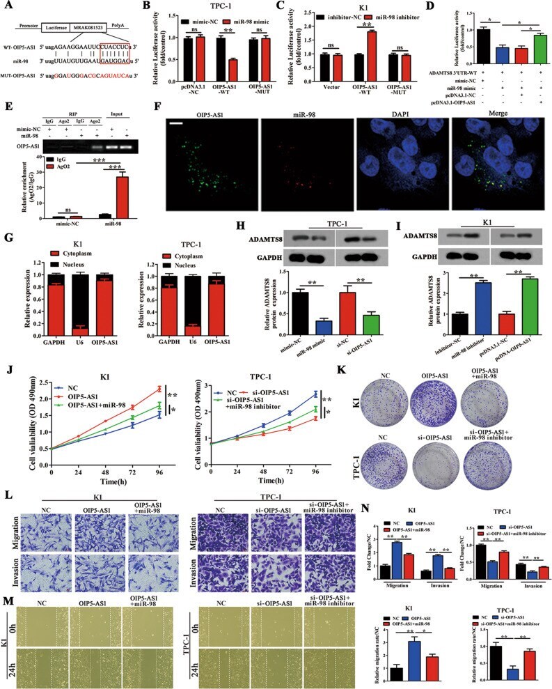
- Fig. 4 OIP5-AS1 is a target of miR-98 and regulates its expression. A Wild-type (OIP5-AS1-WT) and mutant (OIP5-AS1-MUT) OIP5-AS1 with mutations at the predicted miR-98 binding site ( n = 3). B , C A luciferase reporter vector carrying OIP5-AS1-WT or OIP5-AS1-MUT (or the empty vector) was co-transfected into TPC-1 cells with miR-98 mimic or mimic-NC ( B ) or miR-98 inhibitor or inhibitor-NC ( C ) as indicated. Relative luciferase activity was measured at 48 h after transfection ( n = 3). D The luciferase reporter vector carrying ADAMTS8 3'-UTR-WT was co-transfected into TPC-1 cells with miR-98 mimic, mimic-NC, pcDNA3.1-OIP5-AS1, and pcDNA3.1-NC, alone or in combination as indicated. Relative luciferase activity was measured at 48 h after transfection ( n = 3). E TPC-1 cells were transfected with miR-98 mimic or mimic-NC for 48 h, and association between OIP5-AS1 and miR-98 was assessed by RNA immunoprecipitation assay ( n = 3). F Fluorescence in situ hybridization assay with cells stained for OIP5-AS1 and miR-98 ( n = 3) (Scale bar = 20 mum). G Relative lncRNA OIP5-AS1 expression level in the cytoplasm and nucleus of the K1 cells was determined by qRT-PCR assay ( n = 3). H , I Western blot analysis of ADAMTS8 protein levels following treatment of TPC-1 cells with miR-98 mimic or si-OIP5-AS1, and K1 cells with miR-98 inhibitor or pcDNA3.1-OIP5-AS1. GAPDH was used as an internal control ( n = 3). J , K MTT and colony-forming growth assays were performed to determine the prolifer
- Submitted by
- Invitrogen Antibodies (provider)
- Main image
- Experimental details
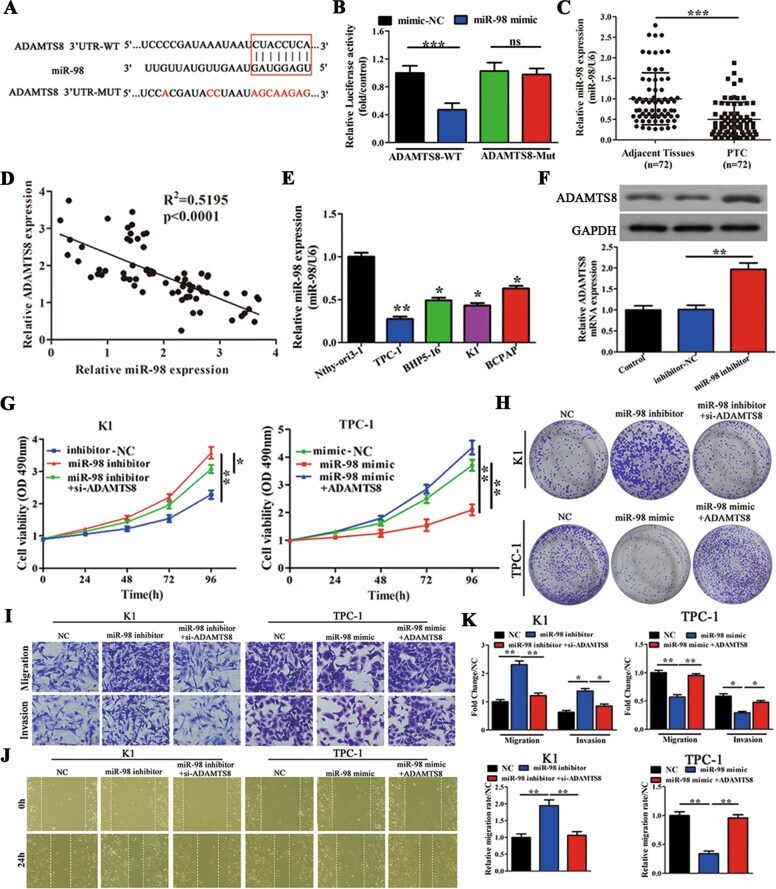
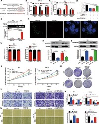
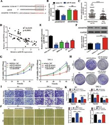
- Fig. 5 ADAMTS8 is a direct target of miR-98 and is involved in miR-98-mediated cell proliferation and migration/invasion. A WT 3'-UTR of ADAMTS8 (ADAMTS8 3'-UTR-WT) and a mutant 3'-UTR of ADAMTS8 with mutations at the predicted miR-98 binding site (ADAMTS8 3'-UTR-MUT) ( n = 3). B A luciferase reporter vector carrying ADAMTS8 3'-UTR-WT or ADAMTS8 3'-UTR-MUT (or the empty vector) was co-transfected with miR-98 mimic or mimic-NC, and relative luciferase activity was measured 48 h post transfection ( n = 3). C qRT-PCR analysis of miR-98 expression in 72 paired PTC tissues and corresponding adjacent tissues. D Pearson correction of miR-98 and ADAMTS8 were analysed ( n = 72). E qRT-PCR analysis of miR-98 expression in four PTC cell lines (TPC-1, BHP5-16, K1, and BCPAP), and in Nthy-ori3-1 cells ( n = 3). F Western blot and qRT-PCR analyses of ADAMTS8 expression levels in K1 cells transfected with inhibitor-NC or miR-98 inhibitor ( n = 3). G , H MTT and colony-forming growth assays were performed to determine the proliferation of K1 and TPC-1 cells ( n = 3). I Transwell assays were performed to determine the migration and invasion capacity of K1 and TPC-1 cells ( n = 3). Scale bars = 50 mum. J Wound healing assays were performed to assess the migratory capacity of K1 and TPC-1 cells ( n = 3). K The migration and invasion abilities and the migratory activity (wound healing) were calculated and compared to the different vectors ( n = 3). * p < 0.05, ** p < 0.01, and *** p < 0.001.