 Explore
 Validate
 Learn
Learn Western blot
Antibody data
- Antibody Data
- Antigen structure
- References [1]
- Comments [0]
- Validations
- Western blot [6]
- Immunocytochemistry [3]
- Immunohistochemistry [1]
Submit
Validation data
Reference
Comment
Report error
- Product number
- PA5-53295 - Provider product page

- Provider
- Invitrogen Antibodies
- Product name
- MST1 (STK4) Polyclonal Antibody
- Antibody type
- Polyclonal
- Antigen
- Recombinant full-length protein
- Description
- Immunogen sequence: IEHDDTLPSQ LGTMVINAED EEEEGTMKRR DETMQPAKPS FLEYFEQKEK ENQINSFGKS VPGPLKNSSD WKIPQDGDYE F Highest antigen sequence identity to the following orthologs: Mouse - 94%, Rat - 94%.
- Reactivity
- Human, Mouse, Rat
- Host
- Rabbit
- Isotype
- IgG
- Vial size
- 100 µL
- Concentration
- 0.1 mg/mL
- Storage
- Store at 4°C short term. For long term storage, store at -20°C, avoiding freeze/thaw cycles.
Submitted references LncRNA SNHG17 knockdown promotes Parkin-dependent mitophagy and reduces apoptosis of podocytes through Mst1.
Guo F, Wang W, Song Y, Wu L, Wang J, Zhao Y, Ma X, Ji H, Liu Y, Li Z, Qin G
Cell cycle (Georgetown, Tex.) 2020 Aug;19(16):1997-2006
Cell cycle (Georgetown, Tex.) 2020 Aug;19(16):1997-2006
No comments: Submit comment
Supportive validation
- Submitted by
- Invitrogen Antibodies (provider)
- Main image
- Experimental details
- Western blot analysis of MST1 in Caco-2 cells transfected with control siRNA, target specific siRNA probe #1, using a MST1 Polyclonal Antibody (Product # PA5-53295). Remaining relative intensity is presented. Loading control: Anti-GAPDH.

- Submitted by
- Invitrogen Antibodies (provider)
- Main image

- Experimental details
- Western blot analysis of MST1 in human cell line CACO-2 using a MST1 Polyclonal Antibody (Product # PA5-53295).

- Submitted by
- Invitrogen Antibodies (provider)
- Main image
- Experimental details
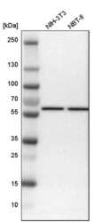
- Western blot analysis of MST1 in mouse cell line NIH-3T3 and rat cell line NBT-II using a MST1 Polyclonal Antibody (Product # PA5-53295).

- Submitted by
- Invitrogen Antibodies (provider)
- Main image

- Experimental details
- Knockout of STK4 was achieved by CRISPR-Cas9 genome editing using LentiArray™ Lentiviral sgRNA (Product # A32042, Assay ID CRISPR879788_LV) and LentiArray Cas9 Lentivirus (Product # A32064). Western blot analysis of STK4 was performed by loading 30 µg of Caco-2 Wild Type (Lane 1), Caco-2 Cas9 (Lane 2) andCaco-2 STK4 KO (Lane 3) whole cell extracts. The samples were electrophoresed using NuPAGE™ Novex™ 4-12% Bis-Tris Protein Gel (Product # NP0322BOX). Resolved proteins were then transferred onto a nitrocellulose membrane (Product # IB23001) by iBlot® 2 Dry Blotting System (Product # IB21001). The blot was probed with a MST1 (STK4) Polyclonal Antibody (Product # PA5-53295, 1:1,000 dilution) and Goat anti-Rabbit IgG (H+L) Superclonal™ Recombinant Secondary Antibody, HRP (Product # A27036, 1:5,000 dilution) using the iBright FL 1000 (Product # A32752). Chemiluminescent detection was performed using Novex® ECL Chemiluminescent Substrate Reagent Kit (Product # WP20005). Loss of signal upon CRISPR mediated knockout (KO) using the LentiArray™ CRISPR product line confirms that antibody is specific to STK4.

- Submitted by
- Invitrogen Antibodies (provider)
- Main image

- Experimental details
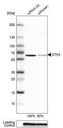
- Knockdown of STK4 was achieved by transfecting Hep G2 with STK4 specific siRNAs (Silencer® select Product # s13571, s13572). Western blot analysis (Fig. a) was performed using Whole cell extracts from the STK4 knockdown cells (lane 3), non-targeting scrambled siRNA transfected cells (lane 2) and untransfected cells (lane 1). The blot was probed with MST1 (STK4) Polyclonal Antibody (Product # PA5-53295, 0.1 µg/mL) and Goat anti-Rabbit IgG (H+L) Superclonal™ Recombinant Secondary Antibody, HRP (Product # A27036, 1:4000 dilution). Densitometric analysis of this western blot is shown in histogram (Fig. b). Decrease in signal upon siRNA mediated knock down confirms that antibody is specific to STK4.

- Submitted by
- Invitrogen Antibodies (provider)
- Main image

- Experimental details
- Western blot was performed using Anti-MST1 (STK4) Polyclonal Antibody (Product # PA5-53295) and a 55 kDa band corresponding to STK4 was observed across cell lines tested. Whole cell extracts (30 µg lysate) of HEK-293 (Lane 1), HeLa (Lane 2), Jurkat (Lane 3), A-431 (Lane 4) and Hep G2 (Lane 5) were electrophoresed using NuPAGE™ 4-12% Bis-Tris Protein Gel (Product # NP0322BOX). Resolved proteins were then transferred onto a Nitrocellulose membrane (Product # IB23001) by iBlot® 2 Dry Blotting System (Product # IB21001). The blot was probed with the primary antibody (0.1 µg/mL) and detected by chemiluminescence with Goat anti-Rabbit IgG (H+L) Superclonal™ Recombinant Secondary Antibody, HRP (Product # A27036, 1:4000 dilution) using the iBright FL 1000 (Product # A32752). Chemiluminescent detection was performed using Novex® ECL Chemiluminescent Substrate Reagent Kit (Product # WP20005).

Supportive validation
- Submitted by
- Invitrogen Antibodies (provider)
- Main image

- Experimental details
- Immunofluorescent staining of MST1 in human cell line CACO-2 shows positivity in nucleus but excluded from the nucleoli. Samples were probed using a MST1 Polyclonal Antibody (Product # PA5-53295).

- Submitted by
- Invitrogen Antibodies (provider)
- Main image
- Experimental details
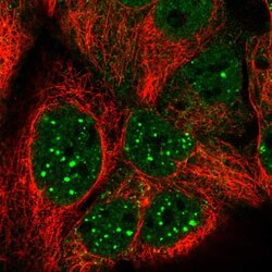
- Immunofluorescent staining of MST1 in human cell line CACO-2 using a MST1 Polyclonal Antibody (Product # PA5-53295) shows localization to nucleoplasm, nuclear bodies and cytosol.

- Submitted by
- Invitrogen Antibodies (provider)
- Main image
- Experimental details
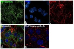
- Immunofluorescence analysis of MST1 was performed using 70% confluent log phase Hep G2 cells. The cells were fixed with 4% paraformaldehyde for 10 minutes, permeabilized with 0.1% Triton™ X-100 for 15 minutes, and blocked with 2% BSA for 45 minutes at room temperature. The cells were labeled with MST1 Polyclonal Antibody (Product # PA5-53295) at 2 µg/mL in 0.1% BSA, incubated at 4 degree celsius overnight and then labeled with Donkey anti-Rabbit IgG (H+L) Highly Cross-Adsorbed Secondary Antibody, Alexa Fluor Plus 488 (Product # A32790), (1:2000), for 45 minutes at room temperature (Panel a: Green). Nuclei (Panel b:Blue) were stained with ProLong™ Diamond Antifade Mountant with DAPI (Product # P36962). F-actin (Panel c: Red) was stained with Rhodamine Phalloidin (Product # R415, 1:300). Panel d represents the merged image showing cytoplasmic localization. Panel e represents control cells with no primary antibody to assess background. The images were captured at 60X magnification.

Supportive validation
- Submitted by
- Invitrogen Antibodies (provider)
- Main image

- Experimental details
- Immunohistochemical staining of MST1 in human lymph node shows strong cytoplasmic positivity in cells outside reaction centra. Samples were probed using a MST1 Polyclonal Antibody (Product # PA5-53295).
