 Explore
 Validate
 Learn
Learn Western blot
 Immunocytochemistry
 Immunohistochemistry
Antibody data
- Antibody Data
- Antigen structure
- References [2]
- Comments [0]
- Validations
- Western blot [1]
- Immunocytochemistry [1]
Submit
Validation data
Reference
Comment
Report error
- Product number
- HPA015270 - Provider product page

- Provider
- Atlas Antibodies
- Proper citation
- Atlas Antibodies Cat#HPA015270, RRID:AB_1854150
- Product name
- Anti-STK4
- Antibody type
- Polyclonal
- Description
- Polyclonal Antibody against Human STK4, Gene description: serine/threonine kinase 4, Alternative Gene Names: KRS2, MST1, YSK3, Validated applications: WB, IHC, ICC, Uniprot ID: Q13043, Storage: Store at +4°C for short term storage. Long time storage is recommended at -20°C.
- Reactivity
- Human, Mouse, Rat
- Host
- Rabbit
- Conjugate
- Unconjugated
- Isotype
- IgG
- Vial size
- 100 µl
- Concentration
- 0.1 mg/ml
- Storage
- Store at +4°C for short term storage. Long time storage is recommended at -20°C.
- Handling
- The antibody solution should be gently mixed before use.
Submitted references STK4 protein expression pattern follows different trends in endometrioid and serous endometrial adenocarcinoma upon tumor progression
Identification of MST1/STK4 and SULF1 Proteins as Autoantibody Targets for the Diagnosis of Colorectal Cancer by Using Phage Microarrays
Govorov I, Attarha S, Kovalevska L, Andersson E, Kashuba E, Mints M
Scientific Reports 2022;12(1)
Scientific Reports 2022;12(1)
Identification of MST1/STK4 and SULF1 Proteins as Autoantibody Targets for the Diagnosis of Colorectal Cancer by Using Phage Microarrays
Babel I, Barderas R, Diaz-Uriarte R, Moreno V, Suarez A, Fernandez-Aceñero M, Salazar R, Capellá G, Casal J
Molecular & Cellular Proteomics 2011;10(3):M110.001784
Molecular & Cellular Proteomics 2011;10(3):M110.001784
No comments: Submit comment
Enhanced validation
- Submitted by
- Atlas Antibodies (provider)
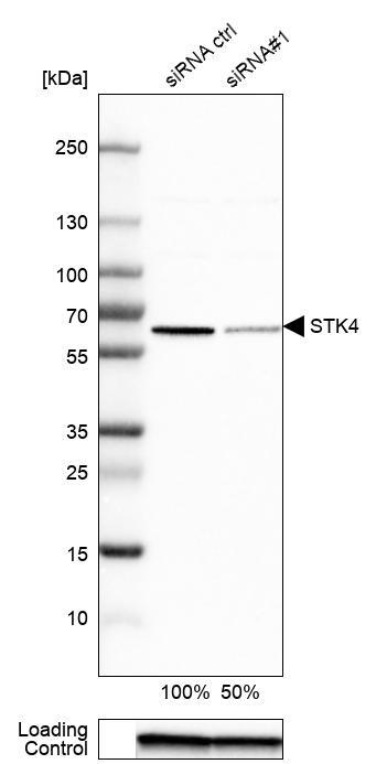
- Enhanced method
- Genetic validation
- Main image
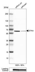
- Experimental details
- Western blot analysis in Caco-2 cells transfected with control siRNA, target specific siRNA probe #1, using Anti-STK4 antibody. Remaining relative intensity is presented. Loading control: Anti-GAPDH.
- Sample type
- Human
- Protocol
- Protocol
Supportive validation
- Submitted by
- Atlas Antibodies (provider)
- Main image
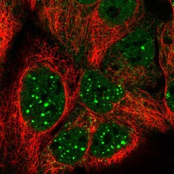
- Experimental details
- Immunofluorescent staining of human cell line CACO-2 shows localization to nucleoplasm, nuclear bodies & cytosol.
- Sample type
- Human
