 Explore
 Validate
 Learn
Learn Flow cytometry
Antibody data
- Antibody Data
- Antigen structure
- References [6]
- Comments [0]
- Validations
- Flow cytometry [1]
Submit
Validation data
Reference
Comment
Report error
- Product number
- 12-9043-41 - Provider product page

- Provider
- Invitrogen Antibodies
- Product name
- Anti-MERTK Monoclonal Antibody (HMER5DS), PE, eBioscience™
- Antibody type
- Monoclonal
- Antigen
- Other
- Description
- Description: This HMER5DS antibody recognizes human MerTK, known also as Mer or c-mer, a 170-210 kDa member of the TAM family of tyrosine kinase receptors that also includes Axl and Tyro3. MerTK is expressed on a subset of anti-inflammatory macrophages and is involved in the removal of apoptotic cells. This process relies on two soluble ligands of Mer,TK Protein S and Gas6, that coat the surface of cells undergoing apoptosis. Upon binding these ligands, MerTK undergoes autophosphorylation at multiple tyrosine residues that activate the PI3K and Akt pathways. This results in the phagocytosis of target cells and also the direct inhibition of TLR-induced production of pro-inflammatory cytokines by the phagocytes. Mer may also function as a putative entry receptor for filoviruses. Deficiency of MerTK causes general autoimmunity, inflammation, and accumulation of apoptotic bodies. MerTK can be released from the cell surface by metalloproteinases, and this process is significantly enhanced when macrophages are stimulated, e.g. with LPS. MerTK is often expressed on malignant cells, and may be implicated in immune evasion. Applications Reported: This HMER5DS antibody has been reported for use in flow cytometric analysis. Applications Tested: This HMER5DS antibody has been pre-titrated and tested by flow cytometric analysis of human monocyte-derived macrophages. This can be used at 5 µL (0.06 µg) per test. A test is defined as the amount (µg) of antibody that will stain a cell sample in a final volume of 100 µL. Cell number should be determined empirically but can range from 10^5 to 10^8 cells/test. Excitation: 488-561 nm; Emission: 578 nm; Laser: Blue Laser, Green Laser, Yellow-Green Laser. Filtration: 0.2 µm post-manufacturing filtered.
- Reactivity
- Human
- Host
- Mouse
- Conjugate
- Yellow dye
- Isotype
- IgG
- Antibody clone number
- HMER5DS
- Vial size
- 25 Tests
- Concentration
- 5 µL/Test
- Storage
- 4° C, store in dark, DO NOT FREEZE!
Submitted references Immune activation caused by vascular oxidation promotes fibrosis and hypertension.
Mer receptor tyrosine kinase mediates both tethering and phagocytosis of apoptotic cells.
Diversification of TAM receptor tyrosine kinase function.
Circulating levels of soluble MER in lupus reflect M2c activation of monocytes/macrophages, autoantibody specificities and disease activity.
TAM receptor function in the retinal pigment epithelium.
The anticoagulation factor protein S and its relative, Gas6, are ligands for the Tyro 3/Axl family of receptor tyrosine kinases.
Wu J, Saleh MA, Kirabo A, Itani HA, Montaniel KR, Xiao L, Chen W, Mernaugh RL, Cai H, Bernstein KE, Goronzy JJ, Weyand CM, Curci JA, Barbaro NR, Moreno H, Davies SS, Roberts LJ 2nd, Madhur MS, Harrison DG
The Journal of clinical investigation 2016 Jan;126(1):50-67
The Journal of clinical investigation 2016 Jan;126(1):50-67
Mer receptor tyrosine kinase mediates both tethering and phagocytosis of apoptotic cells.
Dransfield I, Zagórska A, Lew ED, Michail K, Lemke G
Cell death & disease 2015 Feb 19;6:e1646
Cell death & disease 2015 Feb 19;6:e1646
Diversification of TAM receptor tyrosine kinase function.
Zagórska A, Través PG, Lew ED, Dransfield I, Lemke G
Nature immunology 2014 Oct;15(10):920-8
Nature immunology 2014 Oct;15(10):920-8
Circulating levels of soluble MER in lupus reflect M2c activation of monocytes/macrophages, autoantibody specificities and disease activity.
Zizzo G, Guerrieri J, Dittman LM, Merrill JT, Cohen PL
Arthritis research & therapy 2013;15(6):R212
Arthritis research & therapy 2013;15(6):R212
TAM receptor function in the retinal pigment epithelium.
Prasad D, Rothlin CV, Burrola P, Burstyn-Cohen T, Lu Q, Garcia de Frutos P, Lemke G
Molecular and cellular neurosciences 2006 Sep;33(1):96-108
Molecular and cellular neurosciences 2006 Sep;33(1):96-108
The anticoagulation factor protein S and its relative, Gas6, are ligands for the Tyro 3/Axl family of receptor tyrosine kinases.
Stitt TN, Conn G, Gore M, Lai C, Bruno J, Radziejewski C, Mattsson K, Fisher J, Gies DR, Jones PF
Cell 1995 Feb 24;80(4):661-70
Cell 1995 Feb 24;80(4):661-70
No comments: Submit comment
Supportive validation
- Submitted by
- Invitrogen Antibodies (provider)
- Main image
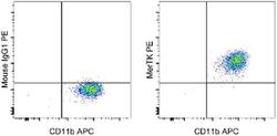
- Experimental details
- Human monocyte-derived macrophages cultured in the presence of Human M-CSF Recombinant Protein (Product # 14-8789-80) and dexamethazone were stained with Anti-Human CD11b APC (Product # 17-0118-42) and Mouse IgG1 K Isotype Control PE (Product # 12-4714-81) (left) or Anti-Human MerTK PE (right). Total viable cells, as determined by Fixable Viability Dye eFluor® 450 (Product # 65-0863-14), were used for analysis.
- Conjugate
- Yellow dye
