Explore
 Validate
 Learn
Learn Western blot
Antibody data
- Antibody Data
- Antigen structure
- References [0]
- Comments [0]
- Validations
- Western blot [3]
- Immunohistochemistry [3]
- Flow cytometry [1]
Submit
Validation data
Reference
Comment
Report error
- Product number
- ATR-033-200UL - Provider product page

- Provider
- Invitrogen Antibodies
- Product name
- MERTK (extracellular) Polyclonal Antibody
- Antibody type
- Polyclonal
- Antigen
- Other
- Reactivity
- Human, Mouse, Rat
- Host
- Rabbit
- Isotype
- IgG
- Vial size
- 200 µL
- Concentration
- 0.8 mg/mL
- Storage
- -20° C, Avoid Freeze/Thaw Cycles
No comments: Submit comment
Supportive validation
- Submitted by
- Invitrogen Antibodies (provider)
- Main image
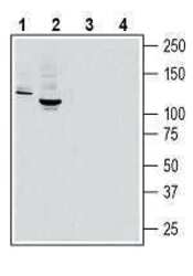
- Experimental details
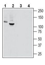
- Western blot analysis of rat brain lysate (lanes 1 and 3) and mouse brain lysate (lanes 2 and 4): - 1, 2. Anti-MERTK (extracellular) Antibody (#ATR-033), (1:400). 3, 4. Anti-MERTK (extracellular) Antibody , preincubated with MERTK (extracellular) Blocking Peptide (#BLP-TR033).

- Submitted by
- Invitrogen Antibodies (provider)
- Main image
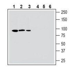
- Experimental details
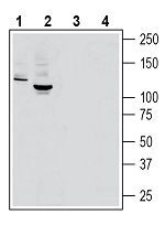
- Western blot analysis human HepG2 hepatocellular carcinoma cell line lysate (lanes 1 and 4), mouse BV-2 microglia cell line lysate (lanes 2 and 5) and human MEG-01 megakaryoblastic leukemia cell line lysate (lanes 3 and 6): - 1-3. Anti-MERTK (extracellular) Antibody (#ATR-033), (1:400). 4-6. Anti-MERTK (extracellular) Antibody , preincubated with MERTK (extracellular) Blocking Peptide (#BLP-TR033).
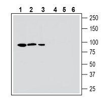
- Submitted by
- Invitrogen Antibodies (provider)
- Main image
- Experimental details
- Western blot analysis of rat brain lysate (lanes 1 and 3) and mouse brain lysate (lanes 2 and 4): - 1, 2. Anti-MERTK (extracellular) Antibody (#ATR-033), (1:400). 3, 4. Anti-MERTK (extracellular) Antibody , preincubated with MERTK (extracellular) Blocking Peptide (#BLP-TR033).
Supportive validation
- Submitted by
- Invitrogen Antibodies (provider)
- Main image
- Experimental details
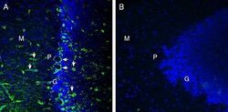
- Expression of MERTK in rat cerebellum - Immunohistochemical staining of perfusion-fixed frozen rat brain sections with Anti-MERTK (extracellular) Antibody (#ATR-033), (1:300), followed by goat anti rabbit conjugated with Alexa 488 (green). A. MERTK immunoreactivity appears in in blood vessel profiles (vertical arrows) in the molecular (M), purkinje (P) and granule (G) layers and in basket profiles (horizontal arrows) around Purkinje soma. B. Pre-incubation of the Antibody with MERTK (extracellular) Blocking Peptide (#BLP-TR033), suppresses staining. Cell nuclei are stained with DAPI (blue). G= Granule layer, P = Purkinje layer, M = Molecular layer.
- Submitted by
- Invitrogen Antibodies (provider)
- Main image

- Experimental details
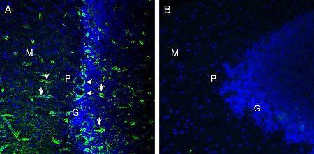
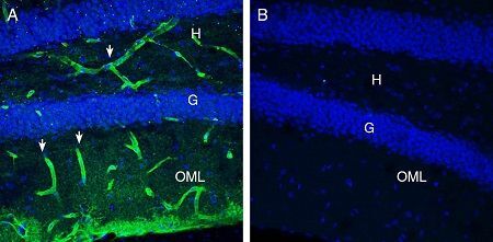
- Expression of MERTK in mouse hippocampus - Immunohistochemical staining of perfusion-fixed frozen mouse brain sections with Anti-MERTK (extracellular) Antibody (#ATR-033), (1:300), followed by goat Anti-rabbit-AlexaFluor-488 (green). A. MERTK staining (green) in the mouse hippocampal dentate gyrus region is detected in blood vessel profiles (arrows) in the hilus (H) and outer molecular layer (OML). B. Pre-incubation of the Antibody with MERTK (extracellular) Blocking Peptide (#BLP-TR033), suppresses staining. Cell nuclei are stained with DAPI (blue). G = Granule layer.
- Submitted by
- Invitrogen Antibodies (provider)
- Main image
- Experimental details
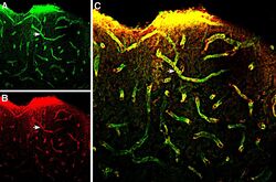
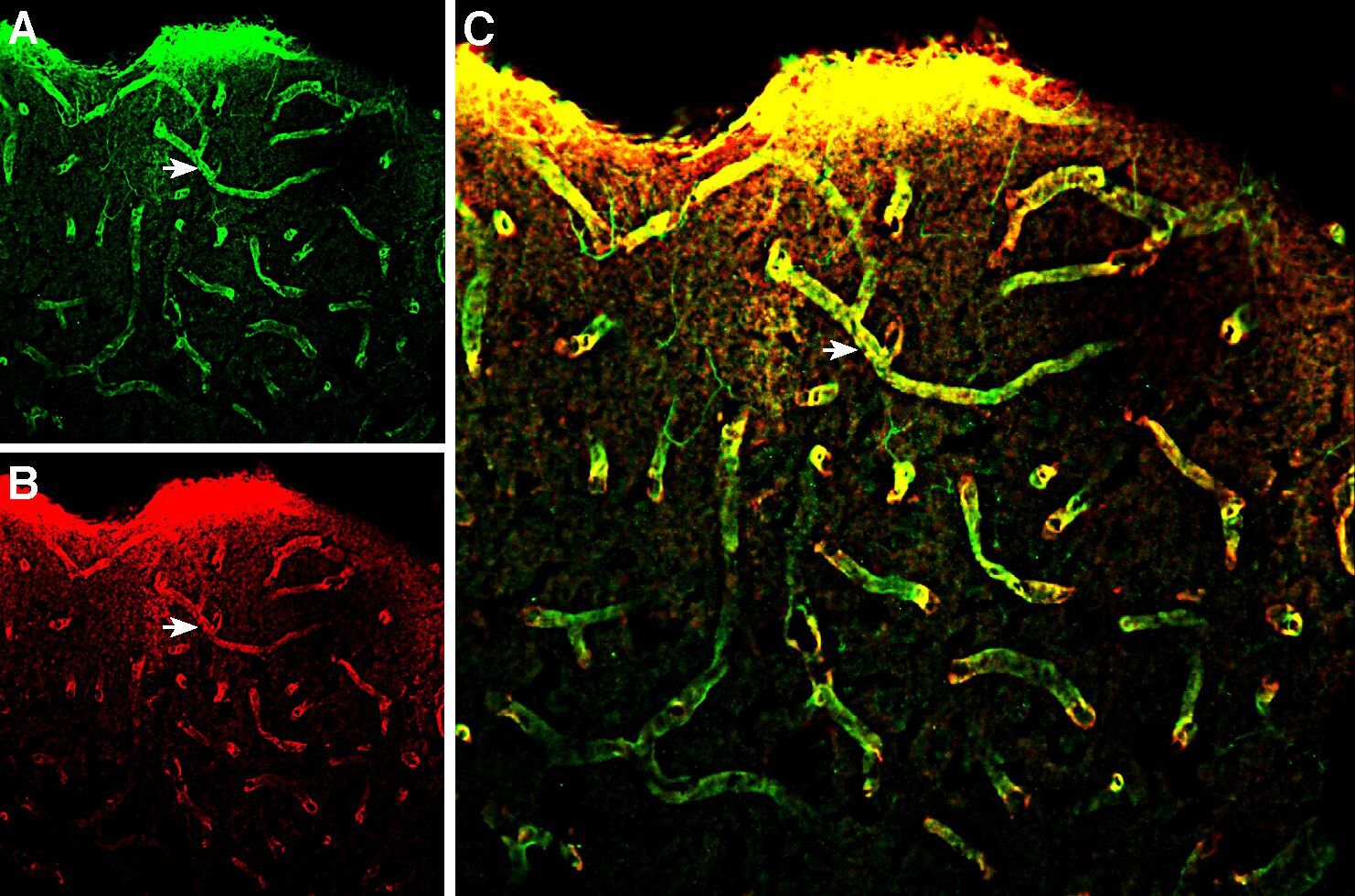
- Multiplex staining of MERTK and Aquaporin 4 in rat parietal cortex. Immunohistochemical staining of perfusion-fixed frozen rat brain sections with Anti-MERTK (extracellular) Antibody (#ATR-033), (1:300), followed by goat Anti-rabbit-AlexaFluor-488 and Guinea Pig Anti-Aquaporin 4 (AQP4) (300-314) Antibody (#AQP-014-GP), (1:300), followed by goat Anti-guinea pig-AlexaFluor-594. A. MERTK immunoreactivity (greens) appears around blood vessels (arrows). B. Aquaporin 4 immunoreactivity (red) appears around blood vessels (arrows). C. Merge of the two images reveals several vessels that express both MERTK and AQP4 (arrows point at examples).
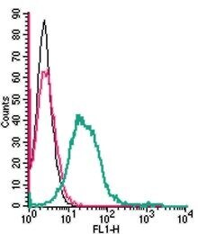
Supportive validation
- Submitted by
- Invitrogen Antibodies (provider)
- Main image

- Experimental details
- Cell surface detection of MERTK in live intact mouse BV-2 microglia cell line: - (black line) cells. (red) Cells + goat- Anti-rabbit-FITC. (green) Cells + Anti-MERTK (extracellular) Antibody (#ATR-033), (5 µg) + goat- Anti-rabbit-FITC.