 Explore
 Validate
 Learn
Learn Western blot
 Immunocytochemistry
Antibody data
- Antibody Data
- Antigen structure
- References [1]
- Comments [0]
- Validations
- Immunocytochemistry [2]
- Immunohistochemistry [1]
- Flow cytometry [3]
Submit
Validation data
Reference
Comment
Report error
- Product number
- PA5-47030 - Provider product page

- Provider
- Invitrogen Antibodies
- Product name
- TROP2 Polyclonal Antibody
- Antibody type
- Polyclonal
- Antigen
- Recombinant full-length protein
- Description
- In direct ELISAs and Western blots, less than 1% cross-reactivity with recombinant human (rh) MCAM, rhNCAM-L1, and rhBCAM is observed. Reconstitute at 0.2 mg/mL in sterile PBS.
- Reactivity
- Human
- Host
- Goat
- Isotype
- IgG
- Vial size
- 100 μg
- Concentration
- 0.2 mg/mL
- Storage
- -20°C, Avoid Freeze/Thaw Cycles
Submitted references SARS-CoV-2 can infect and propagate in human placenta explants.
Fahmi A, Brügger M, Démoulins T, Zumkehr B, Oliveira Esteves BI, Bracher L, Wotzkow C, Blank F, Thiel V, Baud D, Alves MP
Cell reports. Medicine 2021 Dec 21;2(12):100456
Cell reports. Medicine 2021 Dec 21;2(12):100456
No comments: Submit comment
Supportive validation
- Submitted by
- Invitrogen Antibodies (provider)
- Main image
- Experimental details
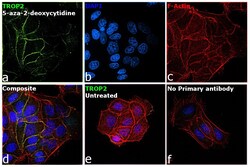
- Immunofluorescence analysis of TROP2 was performed using 70% confluent log phase MCF-7 cells treated with 5-aza-2-deoxycytidine (2.5 µM for 48 hours). The cells were fixed with 4% paraformaldehyde for 10 minutes, permeabilized with 0.1% Triton™ X-100 for 15 minutes, and blocked with 2% BSA for 1 hour at room temperature. The cells were labeled with TROP2 Polyclonal Antibody (Product # PA5-47030) at 2 µg/mL in 0.1% BSA, incubated at 4 degree Celsius overnight and then labeled with Rabbit anti-Goat IgG (H+L), Superclonal™ Recombinant Secondary Antibody, Alexa Fluor 488 (Product # A27012) at a dilution of 1:2000 for 45 minutes at room temperature (Panel a: green). Nuclei (Panel b: blue) were stained with SlowFade® Gold Antifade Mountant with DAPI (Product # S36938). F-actin (Panel c: red) was stained with Rhodamine Phalloidin (Product # R415, 1:300). Panel d represents the merged image showing increased TROP2 expression and localization to plasma membrane upon treatment with 5-aza-2-deoxycytidine. Panel e shows untreated cells with lower expression of TROP2. Panel f represents control cells with no primary antibody to assess background. The images were captured at 60X magnification.
- Submitted by
- Invitrogen Antibodies (provider)
- Main image

- Experimental details
- Immunofluorescence analysis of TROP2 was performed using 70% confluent log phase MCF-7 cells treated with 5-aza-2-deoxycytidine (2.5 µM for 48 hours). The cells were fixed with 4% paraformaldehyde for 10 minutes, permeabilized with 0.1% Triton™ X-100 for 15 minutes, and blocked with 2% BSA for 1 hour at room temperature. The cells were labeled with TROP2 Polyclonal Antibody (Product # PA5-47030) at 2 µg/mL in 0.1% BSA, incubated at 4 degree Celsius overnight and then labeled with Rabbit anti-Goat IgG Heavy Chain, Superclonal™ Recombinant Secondary Antibody, Alexa Fluor 488 (Product # A27012) at a dilution of 1:2000 for 45 minutes at room temperature (Panel a: green). Nuclei (Panel b: blue) were stained with SlowFade® Gold Antifade Mountant with DAPI (Product # S36938). F-actin (Panel c: red) was stained with Rhodamine Phalloidin (Product # R415, 1:300). Panel d represents the merged image showing increased TROP2 expression and localization to plasma membrane upon treatment with 5-aza-2-deoxycytidine. Panel e shows untreated cells with lower expression of TROP2. Panel f represents control cells with no primary antibody to assess background. The images were captured at 60X magnification.

Supportive validation
- Submitted by
- Invitrogen Antibodies (provider)
- Main image
- Experimental details
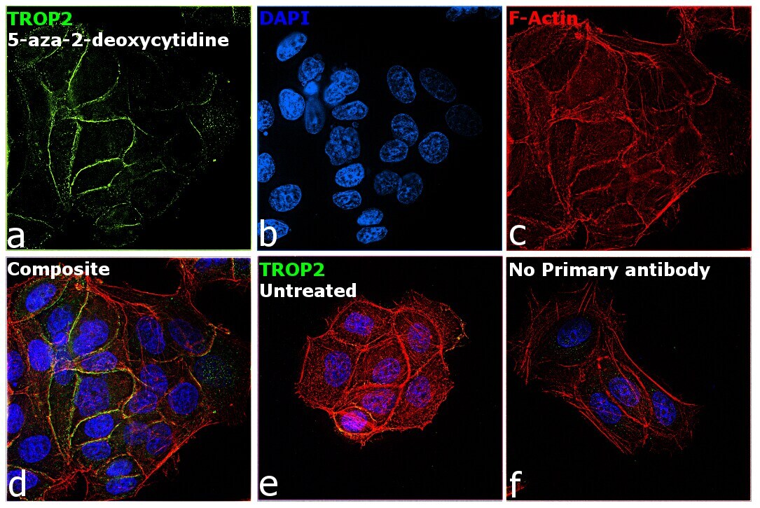
- Immunohistochemical analysis of TROP2 in immersion fixed paraffin-embedded sections of human brain (frontal cortex). Samples were incubated in TROP2 polyclonal antibody (Product # PA5-47030) using a dilution of 10 µg/mL overnight at 4 °C. Before incubation with the primary antibody tissue was subjected to heat-induced epitope retrieval using Antigen Retrieval Reagent-Basic . Tissue was stained using the Anti-Goat HRP-DAB Cell & Tissue Staining Kit (brown) and counterstained with hematoxylin (blue).
Supportive validation
- Submitted by
- Invitrogen Antibodies (provider)
- Main image
- Experimental details
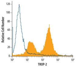
- Flow cytometric analysis of PC-3 human prostate cancer cell line was stained with Goat Anti-human TROP-2 Antigen Affinity-purified Polyclonal Antibody (Product # PA5-47030, filled histogram) or control antibodyopen histogram), followed by Phycoerythrin-conjugated Anti-Goat IgG Secondary Antibody.

- Submitted by
- Invitrogen Antibodies (provider)
- Main image
- Experimental details
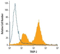
- Flow cytometry of TROP2 in PC‚3 human prostate cancer cell line. Samples were incubated in TROP2 polyclonal antibody (Product # PA5-47030) or control antibody followed by Phycoerythrin-conjugated Anti-Goat IgG Secondary Antibody.

- Submitted by
- Invitrogen Antibodies (provider)
- Main image

- Experimental details
- Flow cytometry of TROP2 in PC‚3 human prostate cancer cell line. Samples were incubated in TROP2 polyclonal antibody (Product # PA5-47030) or control antibody followed by Phycoerythrin-conjugated Anti-Goat IgG Secondary Antibody.
