 Explore
 Validate
 Learn
Learn Western blot
 Immunocytochemistry
Antibody data
- Antibody Data
- Antigen structure
- References [1]
- Comments [0]
- Validations
- Immunocytochemistry [2]
- Immunohistochemistry [1]
- Flow cytometry [3]
Submit
Validation data
Reference
Comment
Report error
- Product number
- PA5-47074 - Provider product page

- Provider
- Invitrogen Antibodies
- Product name
- TROP2 Polyclonal Antibody
- Antibody type
- Polyclonal
- Antigen
- Recombinant full-length protein
- Description
- In direct ELISAs, approximately 30% cross-reactivity with recombinant human TROP-2 is observed. Reconstitute at 0.2 mg/mL in sterile PBS.
- Reactivity
- Human, Mouse, Rat
- Host
- Goat
- Isotype
- IgG
- Vial size
- 100 μg
- Concentration
- 0.2 mg/mL
- Storage
- -20°C, Avoid Freeze/Thaw Cycles
Submitted references Serotonin transporter protects the placental cells against apoptosis in caspase 3-independent pathway.
Hadden C, Fahmi T, Cooper A, Savenka AV, Lupashin VV, Roberts DJ, Maroteaux L, Hauguel-de Mouzon S, Kilic F
Journal of cellular physiology 2017 Dec;232(12):3520-3529
Journal of cellular physiology 2017 Dec;232(12):3520-3529
No comments: Submit comment
Supportive validation
- Submitted by
- Invitrogen Antibodies (provider)
- Main image

- Experimental details
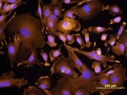
- Immunocytochemistry analysis of TROP2 in immersion fixed XB2 mouse teratoma keratinocyte cell line. Samples were incubated in TROP2 polyclonal antibody (Product # PA5-47074) using a dilution of 10 µg/mL for 3 hours at room temperature followed by NorthernLights™ 557-conjugated Anti-Goat IgG Secondary Antibody (yellow) and counterstained with DAPI (blue).

- Submitted by
- Invitrogen Antibodies (provider)
- Main image
- Experimental details
- Immunocytochemistry analysis of TROP2 in immersion fixed XB2 mouse teratoma keratinocyte cell line. Samples were incubated in TROP2 polyclonal antibody (Product # PA5-47074) using a dilution of 10 µg/mL for 3 hours at room temperature followed by NorthernLights™ 557-conjugated Anti-Goat IgG Secondary Antibody (yellow) and counterstained with DAPI (blue).

Supportive validation
- Submitted by
- Invitrogen Antibodies (provider)
- Main image
- Experimental details
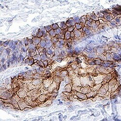
- Immunohistochemical analysis of TROP2 in immersion fixed paraffin-embedded sections of mouse skin. Samples were incubated with TROP2 polyclonal antibody (Product # PA5-47074) using a dilution of 3 µg/mL for 1 hour at room temperature followed by Anti-Goat IgG VisUCyte™ HRP Polymer Antibody. Tissue was stained using DAB (brown) and counterstained with hematoxylin (blue). Specific staining was localized to cell membranes in keratinocytes.

Supportive validation
- Submitted by
- Invitrogen Antibodies (provider)
- Main image

- Experimental details
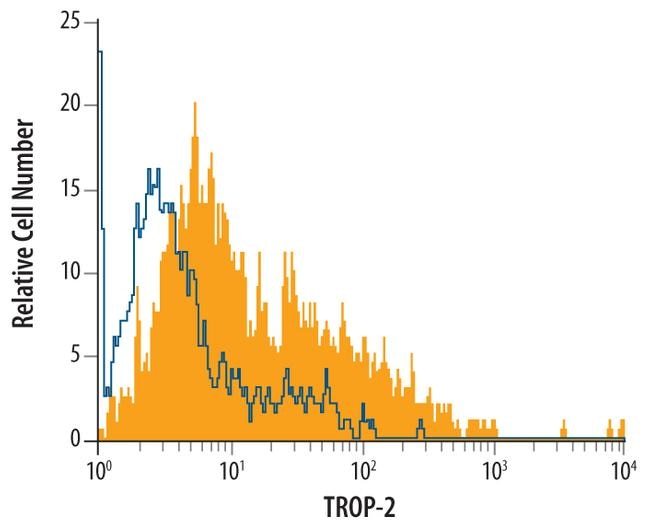
- Flow cytometric analysis of XB2 mouse teratoma keratinocyte cell line was stained with mouse TROP-2 Antigen Affinity-purified Polyclonal Antibody (Product # PA5-47074, filled histogram) or isotype control antibodyopen histogram), followed by Allophycocyanin-conjugated Anti-Goat IgG Secondary Antibody.
- Submitted by
- Invitrogen Antibodies (provider)
- Main image
- Experimental details
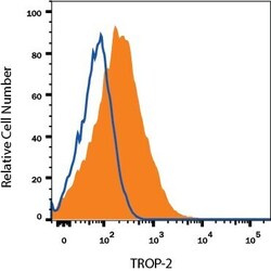
- Flow cytometry of TROP2 in mIMCD-3 mouse epithelial cell line. Samples were incubated in TROP2 polyclonal antibody (Product # PA5-47074) or isotype control antibody followed by Allophycocyanin-conjugated Anti-Goat IgG Secondary Antibody.

- Submitted by
- Invitrogen Antibodies (provider)
- Main image

- Experimental details
- Flow cytometry of TROP2 in mIMCD-3 mouse epithelial cell line. Samples were incubated in TROP2 polyclonal antibody (Product # PA5-47074) or isotype control antibody followed by Allophycocyanin-conjugated Anti-Goat IgG Secondary Antibody.
