Explore
 Validate
 Learn
Learn Immunocytochemistry
Antibody data
- Antibody Data
- Antigen structure
- References [2]
- Comments [0]
- Validations
- Immunocytochemistry [2]
- Immunohistochemistry [6]
- Other assay [1]
Submit
Validation data
Reference
Comment
Report error
- Product number
- PA5-63757 - Provider product page

- Provider
- Invitrogen Antibodies
- Product name
- IYD Polyclonal Antibody
- Antibody type
- Polyclonal
- Antigen
- Recombinant protein fragment
- Description
- Immunogen sequence: ADRSMEKKKG EPRTRAEARP WVDEDLKDSS DLHQAEEDAD EWQESEENVE HIPFSHNHYP EK Highest antigen sequence identity to the following orthologs: Mouse - 66%, Rat - 65%.
- Reactivity
- Human
- Host
- Rabbit
- Isotype
- IgG
- Vial size
- 100 μL
- Concentration
- 0.1 mg/mL
- Storage
- Store at 4°C short term. For long term storage, store at -20°C, avoiding freeze/thaw cycles.
Submitted references Salivary ATP13A2 is a potential marker of therapy-induced motor complications and is expressed by inclusions in submandibulary glands in Parkinson ´s disease.
Native α-Synuclein, 3-Nitrotyrosine Proteins, and Patterns of Nitro-α-Synuclein-Immunoreactive Inclusions in Saliva and Submandibulary Gland in Parkinson's Disease.
Fernández-Espejo E, Gavito AL, Suárez J, Tolosa E, Vilas D, Aldecoa I, Berenguer J, Córdoba-Fernández A, Damas-Hermoso F, Rodríguez de Fonseca F
Clinical parkinsonism & related disorders 2022;7:100163
Clinical parkinsonism & related disorders 2022;7:100163
Native α-Synuclein, 3-Nitrotyrosine Proteins, and Patterns of Nitro-α-Synuclein-Immunoreactive Inclusions in Saliva and Submandibulary Gland in Parkinson's Disease.
Fernández-Espejo E, Rodríguez de Fonseca F, Suárez J, Tolosa E, Vilas D, Aldecoa I, Berenguer J, Damas-Hermoso F
Antioxidants (Basel, Switzerland) 2021 May 1;10(5)
Antioxidants (Basel, Switzerland) 2021 May 1;10(5)
No comments: Submit comment
Supportive validation
- Submitted by
- Invitrogen Antibodies (provider)
- Main image
- Experimental details
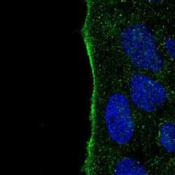
- Immunofluorescent staining of IYD in human cell line CACO-2 using an IYD Polyclonal Antibody (Product # PA5-63757) shows localization to plasma membrane.
- Submitted by
- Invitrogen Antibodies (provider)
- Main image

- Experimental details
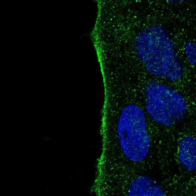
- Immunofluorescent staining of IYD in human cell line CACO-2 using an IYD Polyclonal Antibody (Product # PA5-63757) shows localization to plasma membrane.
Supportive validation
- Submitted by
- Invitrogen Antibodies (provider)
- Main image

- Experimental details
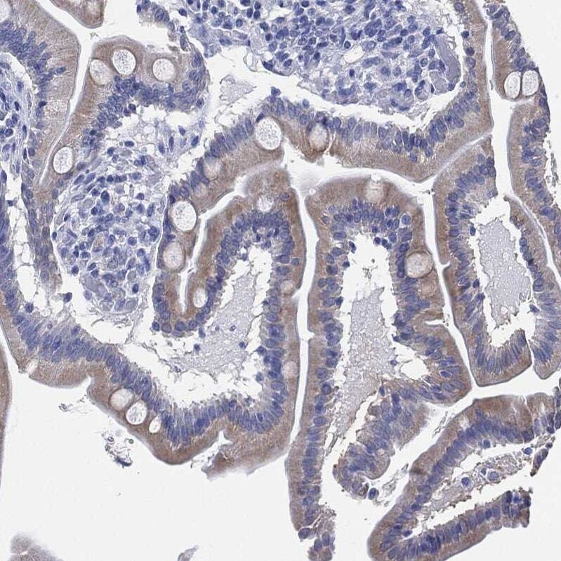
- Immunohistochemical analysis of IYD in human small intestine using IYD Polyclonal Antibody (Product # PA5-63757) shows moderate cytoplasmic positivity in glandular cells.
- Submitted by
- Invitrogen Antibodies (provider)
- Main image
- Experimental details
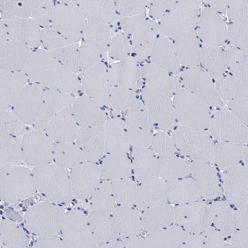
- Immunohistochemical analysis of IYD in human skeletal muscle using IYD Polyclonal Antibody (Product # PA5-63757) shows no positivity in myocytes as expected.
- Submitted by
- Invitrogen Antibodies (provider)
- Main image
- Experimental details
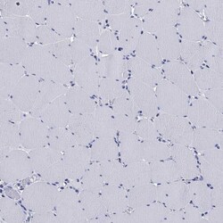
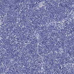
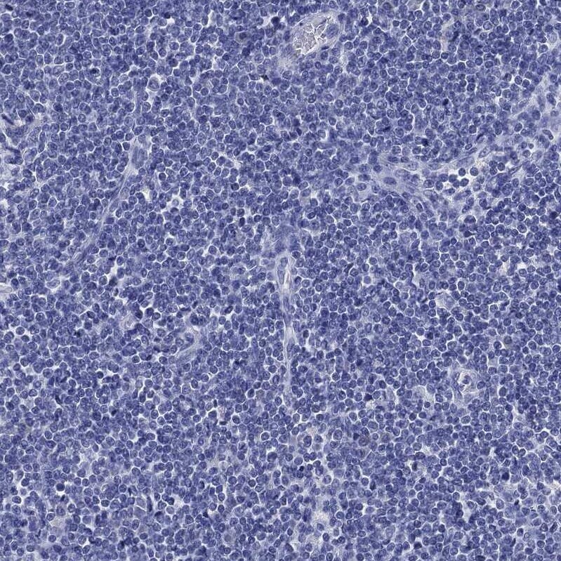
- Immunohistochemical analysis of IYD in human lymph node using IYD Polyclonal Antibody (Product # PA5-63757) shows no positivity in non-germinal center cells as expected.
- Submitted by
- Invitrogen Antibodies (provider)
- Main image
- Experimental details
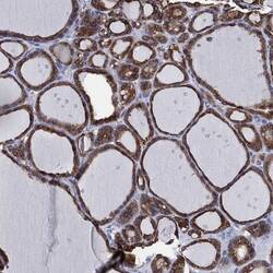
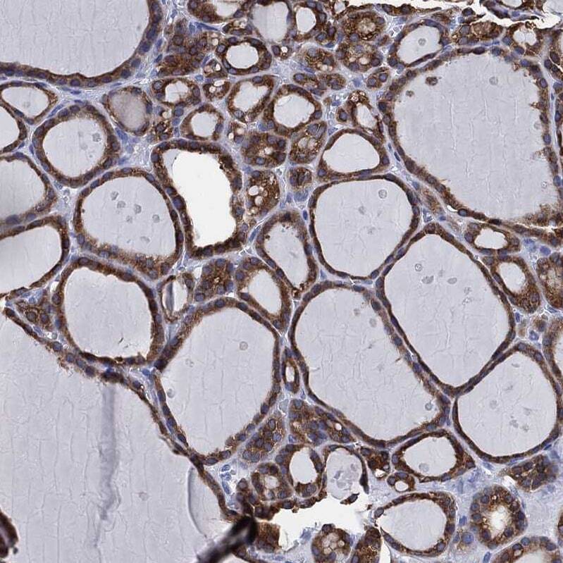
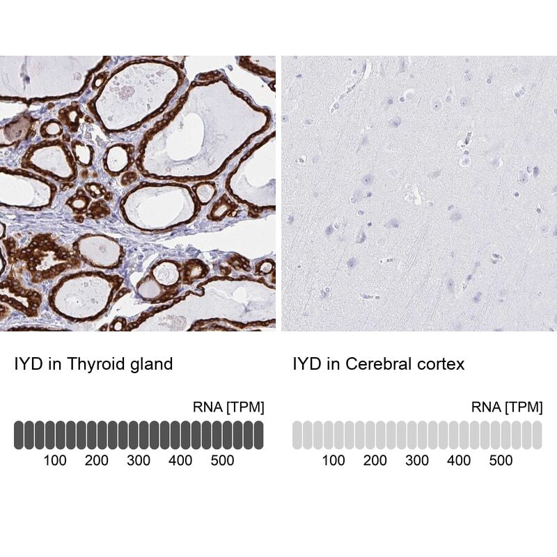
- Immunohistochemical analysis of IYD in human thyroid gland using IYD Polyclonal Antibody (Product # PA5-63757) shows strong cytoplasmic positivity in glandular cells.
- Submitted by
- Invitrogen Antibodies (provider)
- Main image
- Experimental details
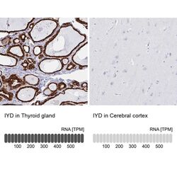
- Immunohistochemical staining of IYD in human thyroid gland and cerebral cortex tissues using IYD Polyclonal Antibody (Product # PA5-63757). Corresponding IYD RNA-seq data are presented for the same tissues.
- Submitted by
- Invitrogen Antibodies (provider)
- Main image
- Experimental details
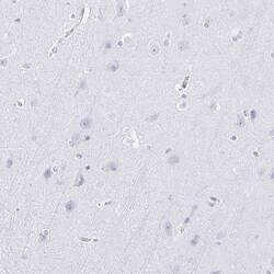
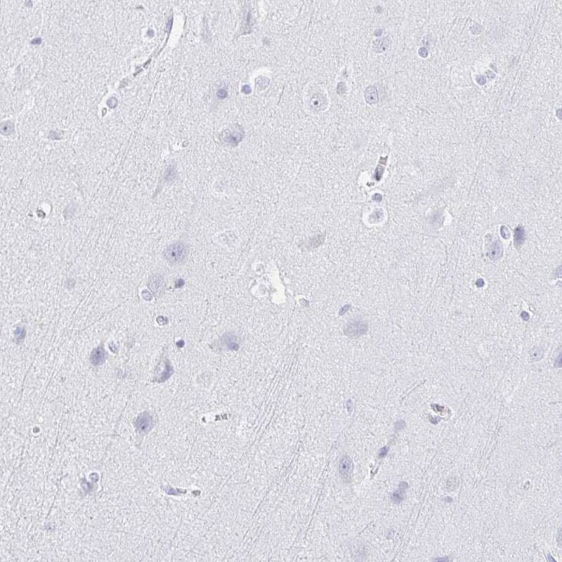
- Immunohistochemical staining of IYD in human cerebral cortex using IYD Polyclonal Antibody (Product # PA5-63757) shows low expression as expected.
Supportive validation
- Submitted by
- Invitrogen Antibodies (provider)
- Main image
- Experimental details
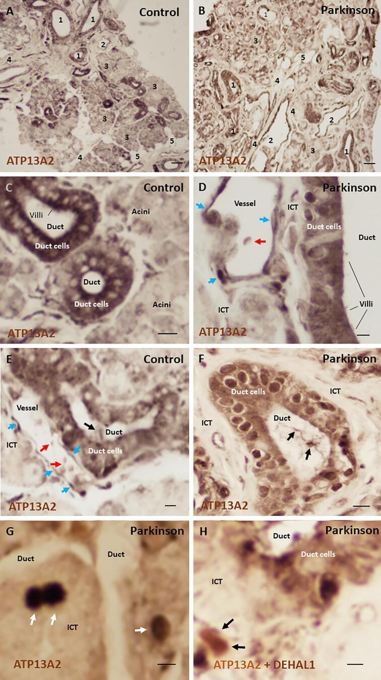
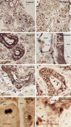
- Representative photomicrographs of submandibulary gland sections in patients with idiopathic Parkinson's disease and controls, after immunostaining against ATP13A2 and DEHAL1. (A, B) Low magnification submandibulary gland sections in a patient and a control participant showing main glandular regions and cell types, as well as intensity of ATP13A2 expression (1, duct cells; 2, vessels with vascular endothelial cells; 3, acinar cells; 4, interlobular connective tissue; 5, adipocytes). (C, D) High-magnification sections of the submandibulary gland showing main cell types that express ATP13A2 in the patients and controls (vascular endothelial cells, blue arrows; red blood cells, red arrows). In C, the difference in staining intensity between excretory duct cells and secretory acinar cells can be observed. In C and D, duct cells show an intracellular gradation of ATP13A2 immunoreactive staining, and the basal region shows less intensity of ATP13A2 expression than the apical region and villi in both the patients and controls. Mild intensity of staining is observed in the interlobular connective tissue. (E-F) ATP13A2-positive deposits inside duct lumen are noticed in the patients and controls (black arrows). These luminal inclusions show amorphous morphology with some fibrils spreading over the lumen. (G-H) Rounded inclusions of 10-20 um in diameter are observed in the interlobular connective tissue of the submandibular gland in patients (black and white arrows). Abbrev. : ATP13A2,