Explore
 Validate
 Learn
Learn Western blot
Antibody data
- Antibody Data
- Antigen structure
- References [0]
- Comments [0]
- Validations
- Western blot [2]
- Immunohistochemistry [3]
- Flow cytometry [1]
Submit
Validation data
Reference
Comment
Report error
- Product number
- F50667 - Provider product page

- Provider
- NSJ Bioreagents
- Product name
- ROR1 Antibody
- Antibody type
- Polyclonal
- Description
- This highly specific ROR1 antibody is suitable for use in Western blot/Flow cytometry/Immunohistochemistry applications with human and mouse samples.
- Reactivity
- Human, Mouse
- Host
- Rabbit
- Conjugate
- Unconjugated
- Vial size
- 0.05 ml, 0.2 ml
- Concentration
- In 1X PBS, pH 7.4, with 0.09% sodium azide
- Storage
- Store at 4oC for up to one month. For long term, aliquot the ROR1 antibody and store frozen at -20oC or colder. Avoid repeated freeze-thaw cycles.
No comments: Submit comment
Supportive validation
- Submitted by
- NSJ Bioreagents (provider)
- Main image
- Experimental details
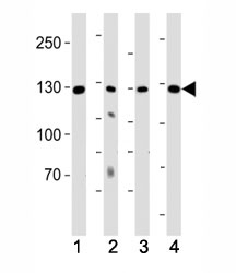
- Western blot analysis of lysate from (1) K562 cell line, (2) human lung, (3) mouse kidney and (4) mouse heart tissue using ROR1 antibody at 1:1000. Predicted molecular weight of ROR1 isoforms: 105 kDa and 130 kDa.

- Submitted by
- NSJ Bioreagents (provider)
- Main image

- Experimental details
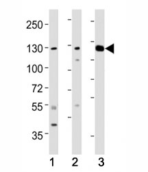
- Western blot testing of ROR1 antibody at 1:2000 dilution. Lane 1: human lung; 2: K562; and 3: mouse kidney lysate; Predicted molecular weight of ROR1 isoforms: 105 kDa and 130 kDa.
Supportive validation
- Submitted by
- NSJ Bioreagents (provider)
- Main image
- Experimental details
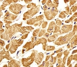
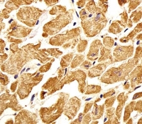
- Immunohistochemical analysis of paraffin-embedded human heart section using ROR1 antibody. Ab was diluted at 1:25 dilution.
- Submitted by
- NSJ Bioreagents (provider)
- Main image
- Experimental details
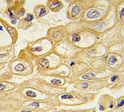
- IHC testing of FFPE human lung carcinoma with ROR1 antibody.

- Submitted by
- NSJ Bioreagents (provider)
- Main image
- Experimental details
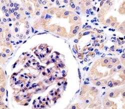
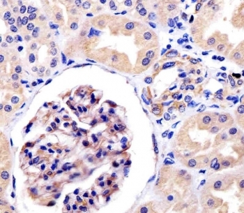
- IHC testing of FFPE human kidney tissue with ROR1 antibody at 1:25 dilution. HIER: steamed antigen retrieval with pH6 citrate buffer.
Supportive validation
- Submitted by
- NSJ Bioreagents (provider)
- Main image
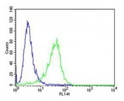
- Experimental details
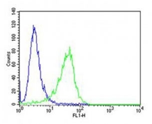
- Flow cytometric analysis of A549 cells using ROR1 antibody (green) compared to an isotype control of rabbit IgG (blue). Ab was diluted at 1:25 dilution. An Alexa Fluor 488 goat anti-rabbit lgG was used as the secondary Ab.