Explore
 Validate
 Learn
Learn Western blot
Antibody data
- Antibody Data
- Antigen structure
- References [12]
- Comments [0]
- Validations
- Western blot [2]
- Flow cytometry [1]
Submit
Validation data
Reference
Comment
Report error
- Product number
- AF2000 - Provider product page

- Provider
- R&D Systems
- Product name
- Human ROR1 Antibody
- Antibody type
- Polyclonal
- Description
- Antigen Affinity-purified. Detects human ROR1 in direct ELISAs and Western blots. In direct ELISAs, less than 5% cross-reactivity with recombinant human ROR2 is observed.
- Reactivity
- Human
- Host
- Goat
- Conjugate
- Unconjugated
- Antigen sequence
Q01973- Isotype
- IgG
- Vial size
- 100 ug
- Concentration
- LYOPH
- Storage
- Use a manual defrost freezer and avoid repeated freeze-thaw cycles. 12 months from date of receipt, -20 to -70 °C as supplied. 1 month, 2 to 8 °C under sterile conditions after reconstitution. 6 months, -20 to -70 °C under sterile conditions after reconstitution.
Submitted references Antibody-drug conjugate T-DM1 treatment for HER2+ breast cancer induces ROR1 and confers resistance through activation of Hippo transcriptional coactivator YAP1.
ROR1-CAVIN3 interaction required for caveolae-dependent endocytosis and pro-survival signaling in lung adenocarcinoma.
A receptor tyrosine kinase ROR1 inhibitor (KAN0439834) induced significant apoptosis of pancreatic cells which was enhanced by erlotinib and ibrutinib.
Maintenance and pharmacologic targeting of ROR1 protein levels via UHRF1 in t(1;19) pre-B-ALL.
ROR1 and ROR2 play distinct and opposing roles in endometrial cancer.
Interaction between ROR1 and MuSK activation complex in myogenic cells.
Silencing ROR1 and ROR2 inhibits invasion and adhesion in an organotypic model of ovarian cancer metastasis.
ROR1 sustains caveolae and survival signalling as a scaffold of cavin-1 and caveolin-1.
Targeting the ROR1 and ROR2 receptors in epithelial ovarian cancer inhibits cell migration and invasion.
Ror1 is a pseudokinase that is crucial for Met-driven tumorigenesis.
The B-cell tumor-associated antigen ROR1 can be targeted with T cells modified to express a ROR1-specific chimeric antigen receptor.
Unique cell surface expression of receptor tyrosine kinase ROR1 in human B-cell chronic lymphocytic leukemia.
Islam SS, Uddin M, Noman ASM, Akter H, Dity NJ, Basiruzzman M, Uddin F, Ahsan J, Annoor S, Alaiya AA, Al-Alwan M, Yeger H, Farhat WA
EBioMedicine 2019 May;43:211-224
EBioMedicine 2019 May;43:211-224
ROR1-CAVIN3 interaction required for caveolae-dependent endocytosis and pro-survival signaling in lung adenocarcinoma.
Yamaguchi T, Hayashi M, Ida L, Yamamoto M, Lu C, Kajino T, Cheng J, Nakatochi M, Isomura H, Yamazaki M, Suzuki M, Fujimoto T, Takahashi T
Oncogene 2019 Jun;38(26):5142-5157
Oncogene 2019 Jun;38(26):5142-5157
A receptor tyrosine kinase ROR1 inhibitor (KAN0439834) induced significant apoptosis of pancreatic cells which was enhanced by erlotinib and ibrutinib.
Daneshmanesh AH, Hojjat-Farsangi M, Ghaderi A, Moshfegh A, Hansson L, Schultz J, Vågberg J, Byström S, Olsson E, Olin T, Österborg A, Mellstedt H
PloS one 2018;13(6):e0198038
PloS one 2018;13(6):e0198038
Maintenance and pharmacologic targeting of ROR1 protein levels via UHRF1 in t(1;19) pre-B-ALL.
Chow M, Gao L, MacManiman JD, Bicocca VT, Chang BH, Alumkal JJ, Tyner JW
Oncogene 2018 Sep;37(38):5221-5232
Oncogene 2018 Sep;37(38):5221-5232
ROR1 and ROR2 play distinct and opposing roles in endometrial cancer.
Henry CE, Llamosas E, Daniels B, Coopes A, Tang K, Ford CE
Gynecologic oncology 2018 Mar;148(3):576-584
Gynecologic oncology 2018 Mar;148(3):576-584
Interaction between ROR1 and MuSK activation complex in myogenic cells.
Karvonen H, Summala K, Niininen W, Barker HR, Ungureanu D
FEBS letters 2018 Feb;592(3):434-445
FEBS letters 2018 Feb;592(3):434-445
Silencing ROR1 and ROR2 inhibits invasion and adhesion in an organotypic model of ovarian cancer metastasis.
Henry C, Hacker N, Ford C
Oncotarget 2017 Dec 22;8(68):112727-112738
Oncotarget 2017 Dec 22;8(68):112727-112738
ROR1 sustains caveolae and survival signalling as a scaffold of cavin-1 and caveolin-1.
Yamaguchi T, Lu C, Ida L, Yanagisawa K, Usukura J, Cheng J, Hotta N, Shimada Y, Isomura H, Suzuki M, Fujimoto T, Takahashi T
Nature communications 2016 Jan 4;7:10060
Nature communications 2016 Jan 4;7:10060
Targeting the ROR1 and ROR2 receptors in epithelial ovarian cancer inhibits cell migration and invasion.
Henry C, Llamosas E, Knipprath-Meszaros A, Schoetzau A, Obermann E, Fuenfschilling M, Caduff R, Fink D, Hacker N, Ward R, Heinzelmann-Schwarz V, Ford C
Oncotarget 2015 Nov 24;6(37):40310-26
Oncotarget 2015 Nov 24;6(37):40310-26
Ror1 is a pseudokinase that is crucial for Met-driven tumorigenesis.
Gentile A, Lazzari L, Benvenuti S, Trusolino L, Comoglio PM
Cancer research 2011 Apr 15;71(8):3132-41
Cancer research 2011 Apr 15;71(8):3132-41
The B-cell tumor-associated antigen ROR1 can be targeted with T cells modified to express a ROR1-specific chimeric antigen receptor.
Hudecek M, Schmitt TM, Baskar S, Lupo-Stanghellini MT, Nishida T, Yamamoto TN, Bleakley M, Turtle CJ, Chang WC, Greisman HA, Wood B, Maloney DG, Jensen MC, Rader C, Riddell SR
Blood 2010 Nov 25;116(22):4532-41
Blood 2010 Nov 25;116(22):4532-41
Unique cell surface expression of receptor tyrosine kinase ROR1 in human B-cell chronic lymphocytic leukemia.
Baskar S, Kwong KY, Hofer T, Levy JM, Kennedy MG, Lee E, Staudt LM, Wilson WH, Wiestner A, Rader C
Clinical cancer research : an official journal of the American Association for Cancer Research 2008 Jan 15;14(2):396-404
Clinical cancer research : an official journal of the American Association for Cancer Research 2008 Jan 15;14(2):396-404
No comments: Submit comment
Supportive validation
- Submitted by
- R&D Systems (provider)
- Main image

- Experimental details
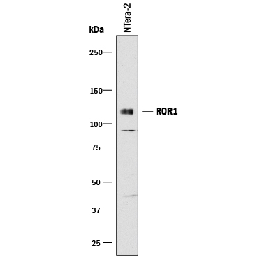
- Detection of ROR1 by Western Blot. Western blot shows lysate of NTera-2 human testicular embryonic carcinoma cell line. PVDF membrane was probed with 1 µg/mL of Goat Anti-Human ROR1 Antigen Affinity-purified Polyclonal Antibody (Catalog # AF2000) followed by HRP-conjugated Anti-Goat IgG Secondary Antibody (Catalog # HAF017). A specific band was detected for ROR1 at approximately 120 kDa (as indicated). This experiment was conducted under reducing conditions and using Immunoblot Buffer Group 1.
- Submitted by
- R&D Systems (provider)
- Main image
- Experimental details
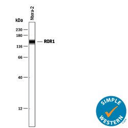
- Detection of Human ROR1 by Simple WesternTM. Simple Western lane view shows lysates of NTera-2 human testicular embryonic carcinoma cell line, loaded at 0.2 mg/mL. A specific band was detected for ROR1 at approximately 143 kDa (as indicated) using 25 µg/mL of Goat Anti-Human ROR1 Antigen Affinity-purified Polyclonal Antibody (Catalog # AF2000) followed by 1:50 dilution of HRP-conjugated Anti-Goat IgG Secondary Antibody (Catalog # HAF109). This experiment was conducted under reducing conditions and using the 12-230 kDa separation system.
Supportive validation
- Submitted by
- R&D Systems (provider)
- Main image

- Experimental details
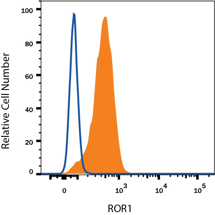
- Detection of ROR1 in MDA-MB-231 Human Cell Line by Flow Cytometry. MDA-MB-231 human breast cancer cell line was stained with Goat Anti-Human ROR1 Antigen Affinity-purified Polyclonal Antibody (Catalog # AF2000, filled histogram) or isotype control antibody (Catalog # AB-108-C, open histogram), followed by Phycoerythrin-conjugated Anti-Goat IgG Secondary Antibody (Catalog # F0107). View our protocol for Staining Membrane-associated Proteins.