Explore
 Validate
 Learn
Learn Western blot
 ELISA
ELISAAntibody data
- Antibody Data
- Antigen structure
- References [0]
- Comments [0]
- Validations
- Western blot [2]
- Immunocytochemistry [5]
- Immunoprecipitation [1]
- Immunohistochemistry [3]
Submit
Validation data
Reference
Comment
Report error
- Product number
- PA5-89139 - Provider product page

- Provider
- Invitrogen Antibodies
- Product name
- DBI Polyclonal Antibody
- Antibody type
- Polyclonal
- Antigen
- Recombinant protein fragment
- Description
- Immunogen sequence: MWGDLWLLPP ASANPGTGTE AEFEKAAEEV RHLKTKPSDE EMLFIYGHYK QATVGDINTE RPGMLDFTGK AKWDAWNELK GTSKEDAMKA YINKVEELKK KYGI; Positive Samples: HT-29, THP-1, MCF-7, NCI-H460, A673, U-937; Cellular Location: Endoplasmic reticulum, Golgi apparatus
- Reactivity
- Human, Mouse, Rat
- Host
- Rabbit
- Isotype
- IgG
- Vial size
- 100 μL
- Concentration
- 0.57 mg/mL
- Storage
- -20°C, Avoid Freeze/Thaw Cycles
No comments: Submit comment
Supportive validation
- Submitted by
- Invitrogen Antibodies (provider)
- Main image
- Experimental details
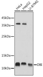
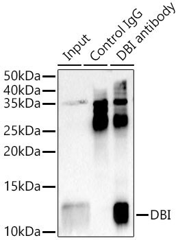
- Western blot analysis of DBI in extracts of various cell lines. Samples were incubated with DBI Polyclonal antibody (Product # PA5-89139) using a dilution of 1:1,000, followed by HRP Goat Anti-Rabbit IgG (H+L) at a dilution of 1:10,000. Lysates/proteins: 25 µg per lane. Blocking buffer: 3% nonfat dry milk in TBST. Detection: ECL Basic Kit. Exposure time: 1s.

- Submitted by
- Invitrogen Antibodies (provider)
- Main image

- Experimental details
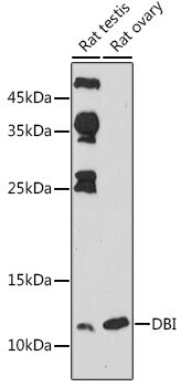
- Western blot analysis of DBI in extracts of various cell lines. Samples were incubated with DBI Polyclonal antibody (Product # PA5-89139) using a dilution of 1:1,000, followed by HRP Goat Anti-Rabbit IgG (H+L) at a dilution of 1:10,000. Lysates/proteins: 25 µg per lane. Blocking buffer: 3% nonfat dry milk in TBST. Detection: ECL Basic Kit. Exposure time: 180s.
Supportive validation
- Submitted by
- Invitrogen Antibodies (provider)
- Main image
- Experimental details
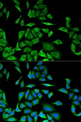
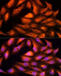
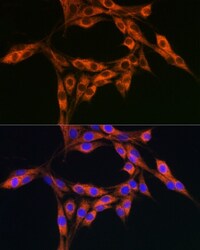
- Immunocytochemistry-Immunofluorescence analysis of DBI was performed in MCF-7 cells using DBI Polyclonal Antibody (Product # PA5-89139). Blue: DAPI for nuclear staining.

- Submitted by
- Invitrogen Antibodies (provider)
- Main image
- Experimental details
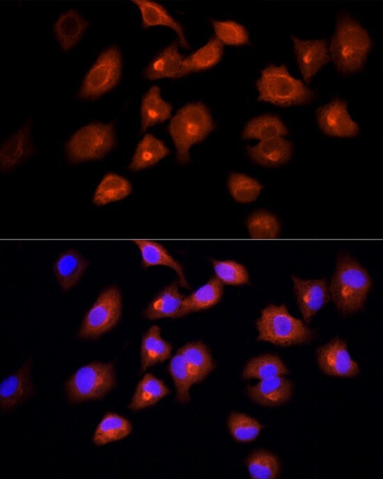
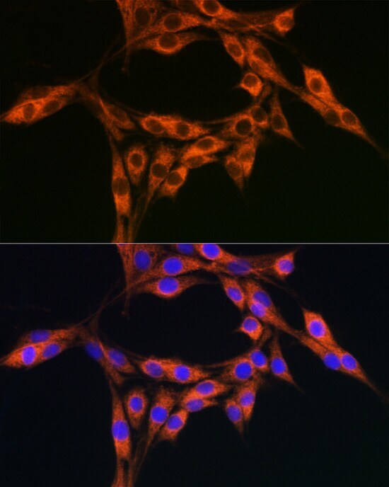
- Immunofluorescence analysis of DBI in U2OS cells. Samples were incubated with DBI Polyclonal antibody (Product # PA5-89139) using a dilution of 1:100 (40x lens). Blue: DAPI for nuclear staining.

- Submitted by
- Invitrogen Antibodies (provider)
- Main image

- Experimental details
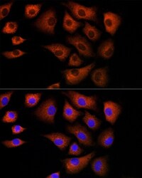
- Immunofluorescence analysis of DBI in A-549 cells. Samples were incubated with DBI Polyclonal antibody (Product # PA5-89139) using a dilution of 1:100 (40x lens). Blue: DAPI for nuclear staining.
- Submitted by
- Invitrogen Antibodies (provider)
- Main image
- Experimental details
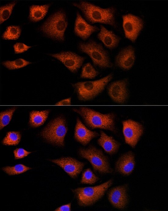
- Immunofluorescence analysis of DBI in NIH/3T3 cells. Samples were incubated with DBI Polyclonal antibody (Product # PA5-89139) using a dilution of 1:100 (40x lens). Blue: DAPI for nuclear staining.
- Submitted by
- Invitrogen Antibodies (provider)
- Main image
- Experimental details
- Immunofluorescence analysis of DBI in PC-12 cells. Samples were incubated with DBI Polyclonal antibody (Product # PA5-89139) using a dilution of 1:100 (40x lens). Blue: DAPI for nuclear staining.
Supportive validation
- Submitted by
- Invitrogen Antibodies (provider)
- Main image

- Experimental details
- Immunoprecipitation of DBI in 300 μg extracts of Hela cells. Samples were precipitated with 3 μg DBI Polyclonal antibody (Product # PA5-89139). Western blot was performed from the immunoprecipitate using DBI Polyclonal antibody (Product # PA5-89139) at a dilution of 1:1,000.
Supportive validation
- Submitted by
- Invitrogen Antibodies (provider)
- Main image
- Experimental details
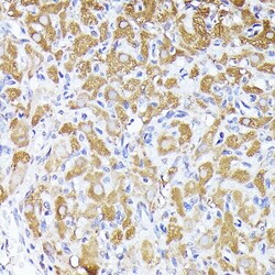
- Immunohistochemistry analysis of DBI in paraffin-embedded rat ovary. Samples were incubated with DBI Polyclonal antibody (Product # PA5-89139) using a dilution of 1:200 (40x lens). Perform high pressure antigen retrieval with 10 mM citrate buffer pH 6.0 before commencing with IHC staining protocol.

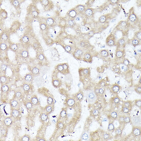
- Submitted by
- Invitrogen Antibodies (provider)
- Main image
- Experimental details
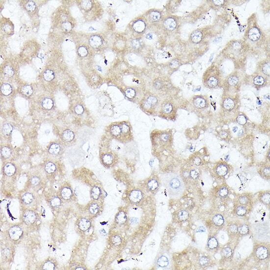
- Immunohistochemistry analysis of DBI in paraffin-embedded rat liver. Samples were incubated with DBI Polyclonal antibody (Product # PA5-89139) using a dilution of 1:200 (40x lens). Perform high pressure antigen retrieval with 10 mM citrate buffer pH 6.0 before commencing with IHC staining protocol.
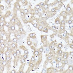
- Submitted by
- Invitrogen Antibodies (provider)
- Main image
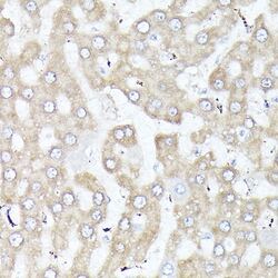
- Experimental details
- Immunohistochemistry analysis of DBI in paraffin-embedded rat liver. Samples were incubated with DBI Polyclonal antibody (Product # PA5-89139) using a dilution of 1:200 (40x lens). Perform high pressure antigen retrieval with 10 mM citrate buffer pH 6.0 before commencing with IHC staining protocol.