 Explore
 Validate
 Learn
Learn Western blot
Antibody data
- Antibody Data
- Antigen structure
- References [0]
- Comments [0]
- Validations
- Western blot [3]
- Immunohistochemistry [4]
Submit
Validation data
Reference
Comment
Report error
- Product number
- LS-C100280 - Provider product page

- Provider
- LSBio
- Proper citation
- LifeSpan Cat#LS-C100280, RRID:AB_2099839
- Product name
- EPHB1 / EPH Receptor B1 Antibody (aa955-984) LS-C100280
- Antibody type
- Polyclonal
- Description
- Ammonium sulfate precipitation
- Reactivity
- Human, Mouse
- Host
- Rabbit
- Storage
- Maintain refrigerated at 2°C to 8°C for up to 6 months. For long term storage store at -20°C.
No comments: Submit comment
Enhanced validation
- Submitted by
- LSBio (provider)
- Enhanced method
- Genetic validation
- Main image
- Experimental details
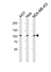
- Western blot of lysates from A431, HeLa, MDA-MB-453 cell line (from left to right), using EPHB1 Antibody (H970). Antibody was diluted at 1:1000 at each lane. A goat anti-rabbit IgG H&L (HRP) at 1:5000 dilution was used as the secondary antibody. Lysates at 35ug per lane.

- Submitted by
- LSBio (provider)
- Enhanced method
- Genetic validation
- Main image
- Experimental details
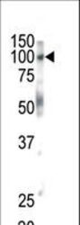
- Western blot of anti-EphB1 antibody in mouse brain tissue. EphB1 (arrow) was detected using purified antibody. Secondary HRP-anti-rabbit was used for signal visualization with chemiluminescence.
- Submitted by
- LSBio (provider)
- Enhanced method
- Genetic validation
- Main image

- Experimental details
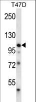
- EPHB1 Antibody (H970) western blot of T47D cell line lysates (35 ug/lane). The EPHB1 antibody detected the EPHB1 protein (arrow).
Enhanced validation
- Submitted by
- LSBio (provider)
- Enhanced method
- Genetic validation
- Main image

- Experimental details
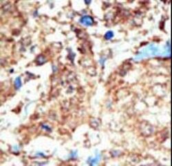
- Formalin-fixed and paraffin-embedded human cancer tissue reacted with the primary antibody, which was peroxidase-conjugated to the secondary antibody, followed by DAB staining. This data demonstrates the use of this antibody for immunohistochemistry; clinical relevance has not been evaluated. BC = breast carcinoma; HC = hepatocarcinoma.

- Submitted by
- LSBio (provider)
- Enhanced method
- Genetic validation
- Main image

- Experimental details
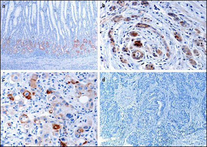
- Immunohistochemical of EphB1 in gastric cancer tissues. a EphB1 protein expressed in normal mucosa at the glandular compartment and in a decreasing gradient from the glandular compartment to the foveolar compartment. b EphB1 protein focally positively stained in well-differentiated gastric cancer cells. c EphB1 protein is focally positive in poorly differentiated gastric cancer cells. d Loss of EphB1 expression in gastric cancer cells.(Provided by Jian-dong Wang,Department of Pathology Nanjing Jinling Hospital/Nanjing University School of Medicine)

- Submitted by
- LSBio (provider)
- Main image

- Experimental details
- Immunohistochemical of EphB1 in gastric cancer tissues. a EphB1 protein expressed in normal mucosa at the glandular compartment and in a decreasing gradient from the glandular compartment to the foveolar compartment. b EphB1 protein focally positively stained in well-differentiated gastric cancer cells. c EphB1 protein is focally positive in poorly differentiated gastric cancer cells. d Loss of EphB1 expression in gastric cancer cells.(Provided by Jian-dong Wang,Department of Pathology Nanjing Jinling Hospital/Nanjing University School of Medicine)
- Submitted by
- LSBio (provider)
- Main image
- Experimental details
- Formalin-fixed and paraffin-embedded human cancer tissue reacted with the primary antibody, which was peroxidase-conjugated to the secondary antibody, followed by DAB staining. This data demonstrates the use of this antibody for immunohistochemistry; clinical relevance has not been evaluated. BC = breast carcinoma; HC = hepatocarcinoma.
