 Explore
 Validate
 Learn
Learn Western blot
Antibody data
- Antibody Data
- Antigen structure
- References [2]
- Comments [0]
- Validations
- Western blot [3]
- Immunohistochemistry [2]
Submit
Validation data
Reference
Comment
Report error
- Product number
- AP7622a - Provider product page

- Provider
- Abcepta
- Proper citation
- Abgent Cat#AP7622a, RRID:AB_2099830
- Product name
- EphB1 Antibody (C-term)
- Antibody type
- Polyclonal
- Antigen
- Synthetic peptide
- Description
- Purified Rabbit Polyclonal Antibody (Pab)
- Reactivity
- Human, Mouse
- Host
- Rabbit
- Isotype
- IgG
- Vial size
- 400 µl
- Storage
- Maintain refrigerated at 2-8°C for up to 6 months. For long term storage store at -20°C in small aliquots to prevent freeze-thaw cycles.
Submitted references EphB1 is underexpressed in poorly differentiated colorectal cancers.
Loss of expression of EphB1 protein in gastric carcinoma associated with invasion and metastasis.
Sheng Z, Wang J, Dong Y, Ma H, Zhou H, Sugimura H, Lu G, Zhou X
Pathobiology : journal of immunopathology, molecular and cellular biology 2008;75(5):274-80
Pathobiology : journal of immunopathology, molecular and cellular biology 2008;75(5):274-80
Loss of expression of EphB1 protein in gastric carcinoma associated with invasion and metastasis.
Wang JD, Dong YC, Sheng Z, Ma HH, Li GL, Wang XL, Lu GM, Sugimura H, Jin J, Zhou XJ
Oncology 2007;73(3-4):238-45
Oncology 2007;73(3-4):238-45
No comments: Submit comment
Supportive validation
- Submitted by
- Abcepta (provider)
- Main image

- Experimental details
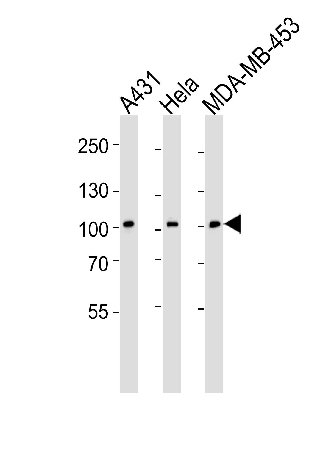
- "Western blot analysis of lysates from A431, Hela, MDA-MB-453 cell line (from left to right), using EPHB1 Antibody (H970)(Cat. #AP7622a). AP7622a was diluted at 1:1000 at each lane. A goat anti-rabbit IgG H&L(HRP) at 1:5000 dilution was used as the secondary antibody. Lysates at 35ug per lane."
- Primary Ab dilution
- 1:1000
- Submitted by
- Abcepta (provider)
- Main image
- Experimental details
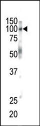
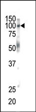
- Western blot analysis of anti-EphB1 Pab (Cat. #AP7622a) in mouse brain tissue. EphB1 (arrow) was detected using purified Pab. Secondary HRP-anti-rabbit was used for signal visualization with chemiluminescence.
- Primary Ab dilution
- 1:1000
- Submitted by
- Abcepta (provider)
- Main image

- Experimental details
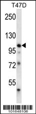
- EPHB1 Antibody (H970) (Cat. #AP7622a) western blot analysis in T47D cell line lysates (35ug/lane).This demonstrates the EPHB1 antibody detected the EPHB1 protein (arrow).
- Primary Ab dilution
- 1:1000
Supportive validation
- Submitted by
- Abcepta (provider)
- Main image
- Experimental details
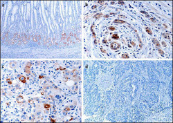
- "Immunohistochemical analysis of EphB1 in gastric cancer tissues. a EphB1 protein expressed in normal mucosa at the glandular compartment and in a decreasing gradient from the glandular compartment to the foveolar compartment. b EphB1 protein focally positively stained in well-differentiated gastric cancer cells. c EphB1 protein is focally positive in poorly differentiated gastric cancer cells. d Loss of EphB1 expression in gastric cancer cells.(Provided by Jian-dong Wang,Department of Pathology Nanjing Jinling Hospital/Nanjing University School of Medicine)"
- Primary Ab dilution
- 1:50~100

- Submitted by
- Abcepta (provider)
- Main image
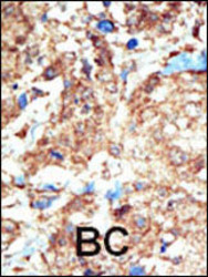
- Experimental details
- "Formalin-fixed and paraffin-embedded human cancer tissue reacted with the primary antibody, which was peroxidase-conjugated to the secondary antibody, followed by DAB staining. This data demonstrates the use of this antibody for immunohistochemistry; clinical relevance has not been evaluated. BC = breast carcinoma; HC = hepatocarcinoma."
- Primary Ab dilution
- 1:50~100
