Explore
 Validate
 Learn
Learn Western blot
 Chromatin Immunoprecipitation
Antibody data
- Antibody Data
- Antigen structure
- References [0]
- Comments [0]
- Validations
- Western blot [3]
- Immunocytochemistry [1]
- Immunohistochemistry [6]
- Other assay [1]
Submit
Validation data
Reference
Comment
Report error
- Product number
- UM500063CF - Provider product page

- Provider
- Invitrogen Antibodies
- Product name
- VBP1 Monoclonal Antibody (UMAB75), UltraMAB™
- Antibody type
- Monoclonal
- Antigen
- Recombinant full-length protein
- Reactivity
- Human
- Host
- Mouse
- Isotype
- IgG
- Antibody clone number
- UMAB75
- Vial size
- 100 µg
- Concentration
- 1 mg/mL
- Storage
- -20° C, Avoid Freeze/Thaw Cycles
No comments: Submit comment
Supportive validation
- Submitted by
- Invitrogen Antibodies (provider)
- Main image
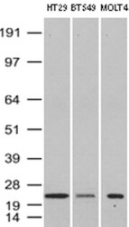
- Experimental details
- Western Blot analysis of HT-29, BT-549 and MOLT-4 cell lysates (35µg) by using anti-VBP1 monoclonal antibody (Clone UMAB75

- Submitted by
- Invitrogen Antibodies (provider)
- Main image
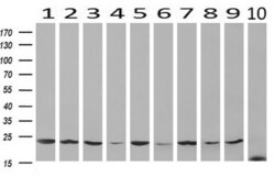
- Experimental details
- Western blot analysis of extracts (10 µg) from 10 Human tissue by using anti-VBP1 monoclonal antibody at 1:200 (1: Testis; 2: Omentum; 3: Uterus; 4: Breast; 5: Brain; 6: Liver; 7: Ovary; 8: Thyroid gland; 9: colon;10: spleen).

- Submitted by
- Invitrogen Antibodies (provider)
- Main image
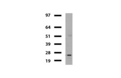
- Experimental details
- Western blot of mouse tissue lysates (20 µg) from Ovary. Primary antibody dilution: 1:500. Secondary antibody dilution: Mouse TrueBlotUltra (1:1000).

Supportive validation
- Submitted by
- Invitrogen Antibodies (provider)
- Main image
- Experimental details
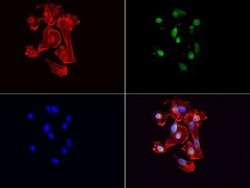
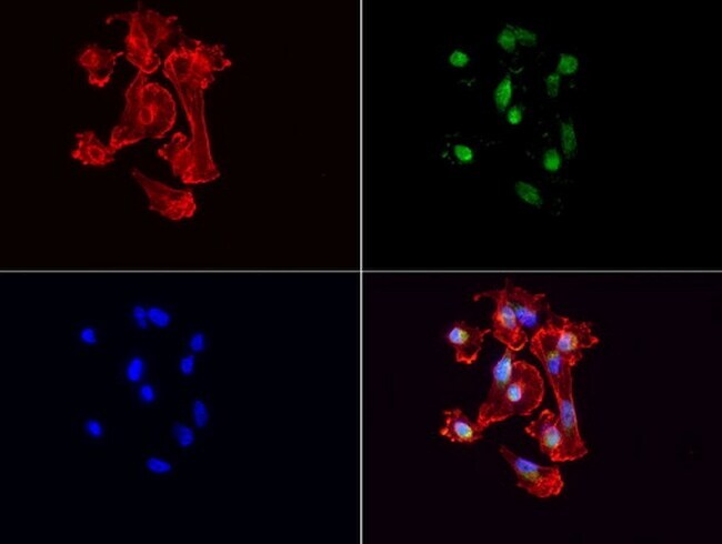
- Immunofluorescent staining of MDA-MB-231 cells using anti-VBP1 mouse monoclonal antibody (UM500063, green, 1:100). Actin filaments were labeled with TRITC-phalloidin (red), and nuclear with DAPI (blue).
Supportive validation
- Submitted by
- Invitrogen Antibodies (provider)
- Main image
- Experimental details
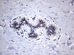
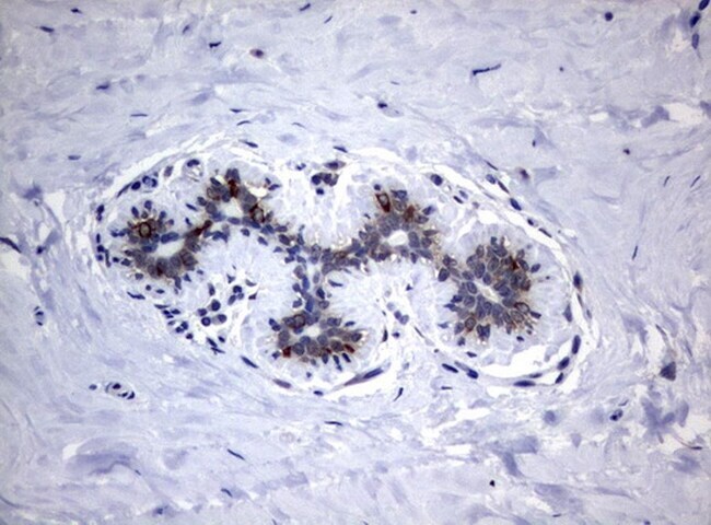
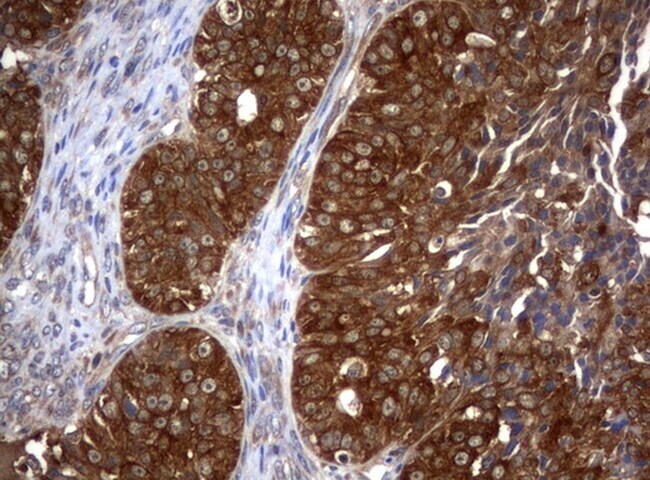
- Immunohistochemical staining of paraffin-embedded human breast tissue using anti-VBP1 mouse monoclonal antibody. (UM500063; heat-induced epitope retrieval by 10mM citric buffer, pH6.0, 120°C for 3min)
- Submitted by
- Invitrogen Antibodies (provider)
- Main image

- Experimental details
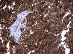
- Immunohistochemical staining of paraffin-embedded Adenocarcinoma of Human ovary tissue using anti-VBP1 mouse monoclonal antibody. (UM500063; heat-induced epitope retrieval by 10mM citric buffer, pH6.0, 120°C for 3min)
- Submitted by
- Invitrogen Antibodies (provider)
- Main image
- Experimental details
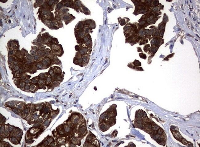
- Immunohistochemical staining of paraffin-embedded Adenocarcinoma of Human endometrium tissue using anti-VBP1 mouse monoclonal antibody. (UM500063; heat-induced epitope retrieval by 10mM citric buffer, pH6.0, 120°C for 3min)

- Submitted by
- Invitrogen Antibodies (provider)
- Main image

- Experimental details
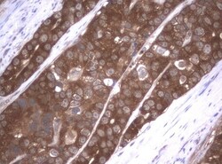
- Immunohistochemical staining of paraffin-embedded Carcinoma of Human bladder tissue using anti-VBP1 mouse monoclonal antibody. (UM500063; heat-induced epitope retrieval by 10mM citric buffer, pH6.0, 120°C for 3min)
- Submitted by
- Invitrogen Antibodies (provider)
- Main image
- Experimental details
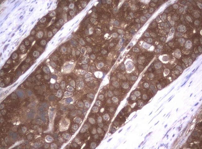
- Immunohistochemical staining of paraffin-embedded Adenocarcinoma of Human colon tissue using anti-VBP1 mouse monoclonal antibody. (UM500063; heat-induced epitope retrieval by 10mM citric buffer, pH6.0, 120°C for 3min)
- Submitted by
- Invitrogen Antibodies (provider)
- Main image

- Experimental details
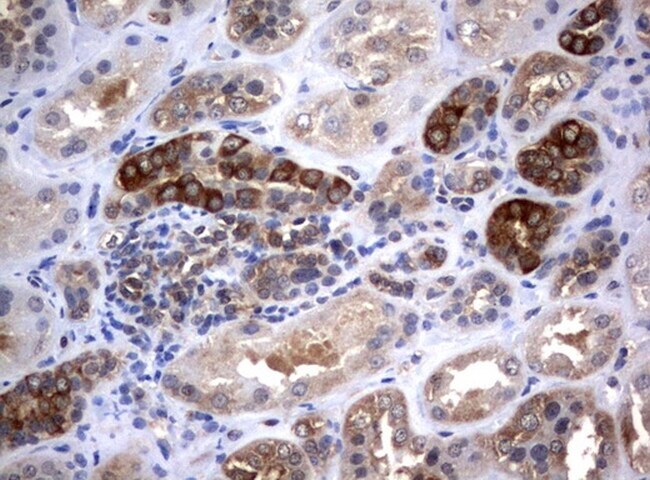
- Immunohistochemical staining of paraffin-embedded human Kidney tissue using anti-VBP1 mouse monoclonal antibody. (UM500063; heat-induced epitope retrieval by 10mM citric buffer, pH6.0, 120°C for 3min)
Supportive validation
- Submitted by
- Invitrogen Antibodies (provider)
- Main image
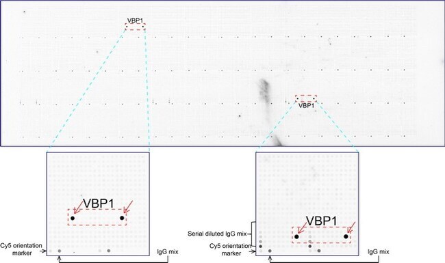
- Experimental details
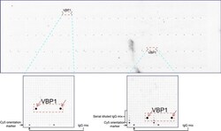
- OriGene overexpression protein microarray chip was immunostained with UltraMAB anti-VBP1 mouse monoclonal antibody (UM500063). The positive reactive proteins are highlighted with two red arrows in the enlarged subarray. All the positive controls spotted in this subarray are also labeled for clarification.