 Explore
 Validate
 Learn
LearnMA5-44978
antibody from Invitrogen Antibodies
Targeting: WLS
C1orf139, EVI, FLJ23091, GPR177, mig-14, MRP
 Western blot
Antibody data
- Antibody Data
- Antigen structure
- References [0]
- Comments [0]
- Validations
- Western blot [2]
- Immunocytochemistry [3]
Submit
Validation data
Reference
Comment
Report error
- Product number
- MA5-44978 - Provider product page

- Provider
- Invitrogen Antibodies
- Product name
- GPR177 Monoclonal Antibody (A7C2)
- Antibody type
- Monoclonal
- Antigen
- Recombinant full-length protein
- Reactivity
- Human, Mouse, Rat
- Host
- Mouse
- Isotype
- IgG
- Antibody clone number
- A7C2
- Vial size
- 100 μL
- Concentration
- 2 mg/mL
- Storage
- Store at 4°C short term. For long term storage, store at -20°C, avoiding freeze/thaw cycles.
No comments: Submit comment
Supportive validation
- Submitted by
- Invitrogen Antibodies (provider)
- Main image
- Experimental details
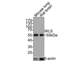
- Western blot analysis of WLS in different lysates (20 µg/Lane). Exposure time: 30 seconds; 10% SDS-PAGE gel. Proteins were transferred to a PVDF membrane and blocked with 5% NFDM/TBST for 1 hour at room temperature. Samples were incubated in WLS Monoclonal antibody (Product # MA5-44978) using a dilution of 1:5,000 in 5% NFDM/TBST at room temperature for 2 hours followed by Goat Anti-Mouse IgG - HRP secondary antibody at a dilution of 1:100,000 for 1 hour at room temperature. Lane 1: Mouse lung tissue lysate; Lane 2: Rat brain tissue lysate. Predicted band size: 62 kDa. Observed band size: 55 kDa.

- Submitted by
- Invitrogen Antibodies (provider)
- Main image

- Experimental details
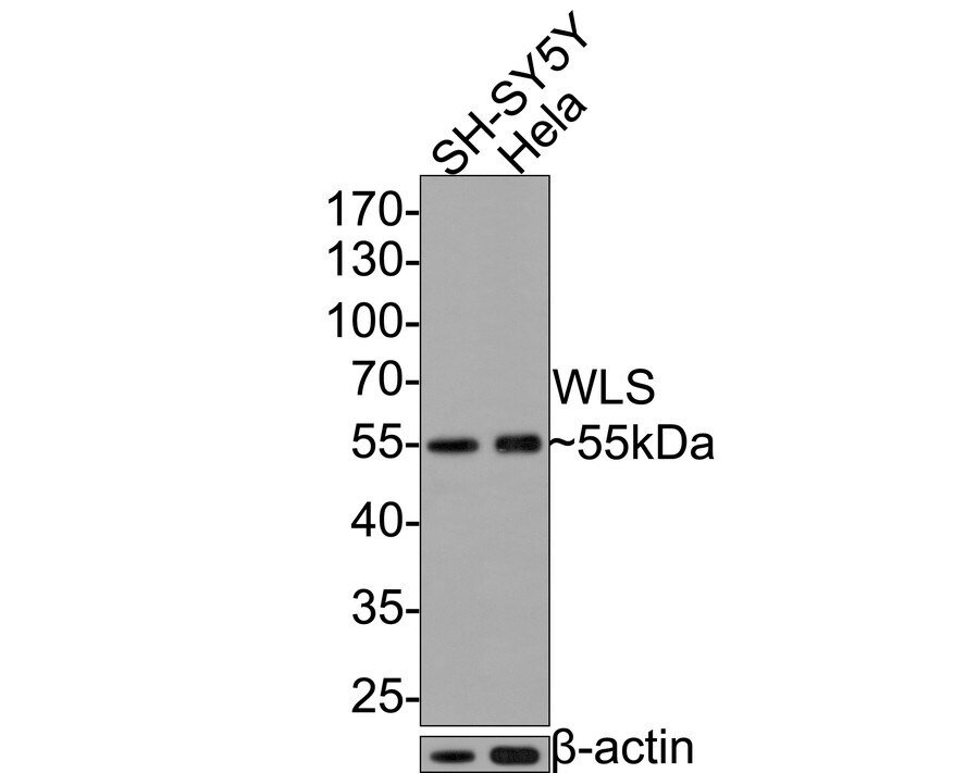
- Western blot analysis of WLS in different lysates (10 µg/Lane). Exposure time: 30 seconds; 10% SDS-PAGE gel. Proteins were transferred to a PVDF membrane and blocked with 5% NFDM/TBST for 1 hour at room temperature. Samples were incubated in WLS Monoclonal antibody (Product # MA5-44978) using a dilution of 1:20,000 in 5% NFDM/TBST at room temperature for 2 hours followed by Goat Anti-Mouse IgG - HRP secondary antibody at a dilution of 1:100,000 for 1 hour at room temperature. Lane 1: SH-SY5Y cell lysate; Lane 2: Hela cell lysate. Predicted band size: 62 kDa. Observed band size: 55 kDa.
Supportive validation
- Submitted by
- Invitrogen Antibodies (provider)
- Main image
- Experimental details
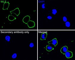
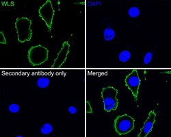
- Immunocytochemistry analysis of WLS in HeLa cells. Cells were fixed in 4% paraformaldehyde for 30 minutes, permeabilized with 0.1% Triton X-100 in PBS for 15 minutes, and then blocked with 2% BSA for 30 minutes at room temperature. Samples were incubated in WLS Monoclonal antibody (Product # MA5-44978) using a dilution of 1:100 in 2% BSA overnight at 4 ℃ followed by Goat Anti-Mouse IgG H&L (iFluor™ 488) secondary antibody at a dilution of 1:1,000. PBS instead of the primary antibody was used as the secondary antibody only control. Nuclear DNA was labelled in blue with DAPI.
- Submitted by
- Invitrogen Antibodies (provider)
- Main image
- Experimental details
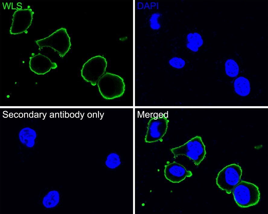
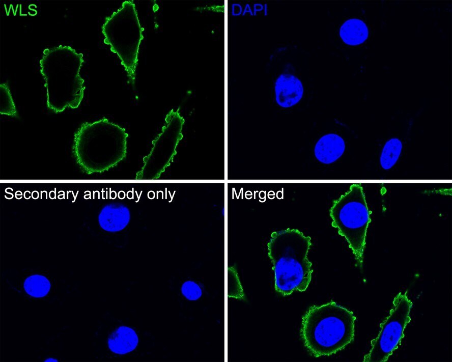
- Immunocytochemistry analysis of WLS in SH-SY5Y cells. Cells were fixed in 4% paraformaldehyde for 30 minutes, permeabilized with 0.1% Triton X-100 in PBS for 15 minutes, and then blocked with 2% BSA for 30 minutes at room temperature. Samples were incubated in WLS Monoclonal antibody (Product # MA5-44978) using a dilution of 1:100 in 2% BSA overnight at 4 ℃ followed by Goat Anti-Mouse IgG H&L (iFluor™ 488) secondary antibody at a dilution of 1:1,000. PBS instead of the primary antibody was used as the secondary antibody only control. Nuclear DNA was labelled in blue with DAPI.
- Submitted by
- Invitrogen Antibodies (provider)
- Main image

- Experimental details
- Immunocytochemistry analysis of WLS in SH-SY5Y cells. Cells were fixed in 4% paraformaldehyde for 30 minutes, permeabilized with 0.1% Triton X-100 in PBS for 15 minutes, and then blocked with 2% BSA for 30 minutes at room temperature. Samples were incubated in WLS Monoclonal antibody (Product # MA5-44978) using a dilution of 1:100 in 2% BSA overnight at 4 ℃ followed by Goat Anti-Mouse IgG H&L (iFluor™ 488) secondary antibody at a dilution of 1:1,000. PBS instead of the primary antibody was used as the secondary antibody only control. Nuclear DNA was labelled in blue with DAPI.
