Explore
 Validate
 Learn
Learn Western blot
 Immunohistochemistry
Antibody data
- Antibody Data
- Antigen structure
- References [1]
- Comments [0]
- Validations
- Immunohistochemistry [1]
- Other assay [1]
Submit
Validation data
Reference
Comment
Report error
- Product number
- PA5-78414 - Provider product page

- Provider
- Invitrogen Antibodies
- Product name
- RPE65 Polyclonal Antibody
- Antibody type
- Polyclonal
- Antigen
- Recombinant full-length protein
- Description
- Positive Control: mouse eye Predicted Reactivity: Rat (93%), Xenopus laevis (83%), Dog (97%), Chicken (89%), Bovine (98%) Store product as a concentrated solution. Centrifuge briefly prior to opening the vial.
- Reactivity
- Human, Mouse
- Host
- Rabbit
- Isotype
- IgG
- Vial size
- 100 μL
- Concentration
- 0.96 mg/mL
- Storage
- Store at 4°C short term. For long term storage, store at -20°C, avoiding freeze/thaw cycles.
Submitted references Evaluation for Retinal Therapy for RPE65 Variation Assessed in hiPSC Retinal Pigment Epithelial Cells.
Nash BM, Loi TH, Fernando M, Sabri A, Robinson J, Cheng A, Eamegdool SS, Farnsworth E, Bennetts B, Grigg JR, Chung SK, Gonzalez-Cordero A, Jamieson RV
Stem cells international 2021;2021:4536382
Stem cells international 2021;2021:4536382
No comments: Submit comment
Supportive validation
- Submitted by
- Invitrogen Antibodies (provider)
- Main image
- Experimental details
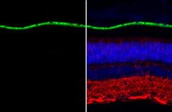
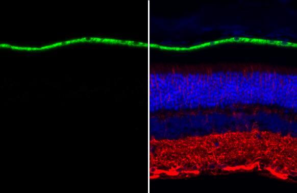
- RPE65 Polyclonal Antibody detects RPE65 protein at retinal pigment epithelium by immunohistochemical analysis. Sample: Paraffin-embedded mouse eye. Green: RPE65 stained by RPE65 Polyclonal Antibody (Product # PA5-78414) diluted at 1:500. Red: beta Tubulin 3/ Tuj1, a neuronal marker, stained by beta Tubulin 3/ Tuj1 antibody [GT11710] diluted at 1:500. Blue: Fluoroshield with DAPI. Antigen Retrieval: Citrate buffer, pH 6.0, 15 min.
Supportive validation
- Submitted by
- Invitrogen Antibodies (provider)
- Main image
- Experimental details
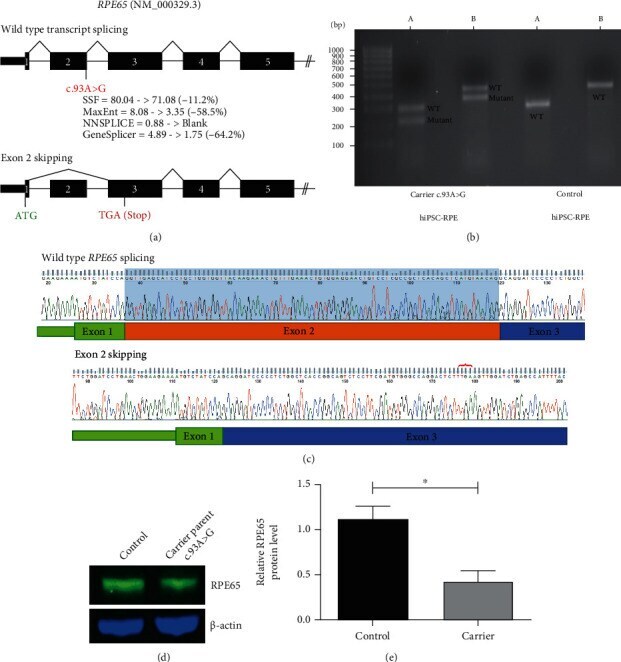
- Figure 4 RNA and protein studies of the RPE65 c.93A>G variant. (a) Schematic of wild-type and mutant transcripts and location of the novel c.93A>G variant are shown. Predicted reduction in splice donor strength due to this variant using Alamut Visual is annotated beneath. The site of the predicted premature stop codon (TGA) is indicated in red on the mutant transcript schematic. (b) Representative agarose gel image of cDNA studies illustrating additional presence of a smaller band consistent with exon skipping in c.93A>G carrier hiPSC-RPE cells compared with control. A: amplicon primers located in 5'UTR and exon 4 (mutant allele = 215 bp; wild-type allele = 297 bp); B: amplicon primers located in 5'UTR and exon 5 (mutant allele = 368 bp; wild-type allele = 450 bp). (c) Purified gel band sequencing of the control and mutant cDNA sequence shows excision of exon 2 from the mutant transcript. Red bracket indicates the location of the premature stop codon introduced. Blue shading indicates normal Exon 2 sequence which is absent from the mutant transcript. (d) Western blot analysis showing RPE65 levels in the control [Left] and parental carrier hiPSC-RPE cells [Right]. (e). Density analysis of RPE65 protein bands shows approximately 50% reduction in RPE65 expression in the parental carrier hiPSC-RPE cells compared with the control (unpaired t -test, n = 3 independent experiments, * P < 0.05).