Explore
 Validate
 Learn
Learn Western blot
 ELISA
ELISA Immunocytochemistry
Antibody data
- Antibody Data
- Antigen structure
- References [2]
- Comments [0]
- Validations
- Immunocytochemistry [2]
- Immunoprecipitation [1]
- Flow cytometry [2]
- Other assay [3]
Submit
Validation data
Reference
Comment
Report error
- Product number
- MA5-29022 - Provider product page

- Provider
- Invitrogen Antibodies
- Product name
- Human Serum Albumin Recombinant Rabbit Monoclonal Antibody (101)
- Antibody type
- Monoclonal
- Antigen
- Other
- Description
- This product is preservative free. It is recommended to add sodium azide to avoid contamination (final concentration 0.05%-0.1%). Recombinant rabbit monoclonal antibodies are produced using in vitro expression systems. The expression systems are developed by cloning in the specific antibody DNA sequences from immunoreactive rabbits. Then, individual clones are screened to select the best candidates for production. The advantages of using recombinant rabbit monoclonal antibodies include: better specificity and sensitivity, lot-to-lot consistency, animal origin-free formulations, and broader immunoreactivity to diverse targets due to larger rabbit immune repertoire. This antibody has specificity for Human Serum Albumin/HSA.
- Reactivity
- Human
- Host
- Rabbit
- Isotype
- IgG
- Antibody clone number
- 101
- Vial size
- 100 μL
- Concentration
- 1 mg/mL
- Storage
- Store at 4°C short term. For long term storage, store at -20°C, avoiding freeze/thaw cycles.
Submitted references Programmable half-life and anti-tumour effects of bispecific T-cell engager-albumin fusions with tuned FcRn affinity.
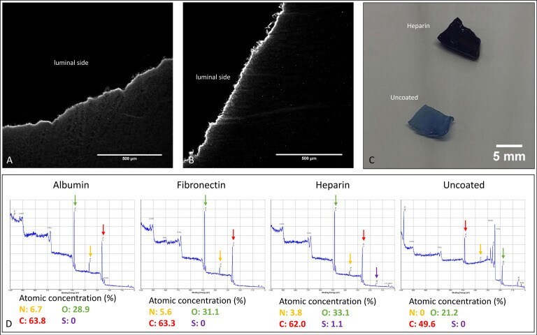
Comparative Evaluation on Impacts of Fibronectin, Heparin-Chitosan, and Albumin Coating of Bacterial Nanocellulose Small-Diameter Vascular Grafts on Endothelialization In Vitro.
Mandrup OA, Ong SC, Lykkemark S, Dinesen A, Rudnik-Jansen I, Dagnæs-Hansen NF, Andersen JT, Alvarez-Vallina L, Howard KA
Communications biology 2021 Mar 8;4(1):310
Communications biology 2021 Mar 8;4(1):310
Comparative Evaluation on Impacts of Fibronectin, Heparin-Chitosan, and Albumin Coating of Bacterial Nanocellulose Small-Diameter Vascular Grafts on Endothelialization In Vitro.
Wacker M, Riedel J, Walles H, Scherner M, Awad G, Varghese S, Schürlein S, Garke B, Veluswamy P, Wippermann J, Hülsmann J
Nanomaterials (Basel, Switzerland) 2021 Jul 29;11(8)
Nanomaterials (Basel, Switzerland) 2021 Jul 29;11(8)
No comments: Submit comment
Supportive validation
- Submitted by
- Invitrogen Antibodies (provider)
- Main image
- Experimental details
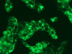
- Immunofluorescence staining of Human HSA in HepG2 cells. Cells were fixed with 4% PFA, permeabilzed with 0.3% Triton X-100 in PBS, blocked with 10% serum, and incubated with Human Serum Albumin Recombinant Rabbit Monoclonal Antibody (101) (Product # MA5-29022, 1:60) at 37°C 1 hour. Then cells were stained with the Alexa Fluor® 488-conjugated Goat Anti-rabbit IgG secondary antibody (green). Positive staining was localized to cytoplasm.

- Submitted by
- Invitrogen Antibodies (provider)
- Main image

- Experimental details
- Immunofluorescence staining of Human HSA in HepG2 cells. Cells were fixed with 4% PFA, permeabilzed with 0.3% Triton X-100 in PBS, blocked with 10% serum, and incubated with Human Serum Albumin Recombinant Rabbit Monoclonal Antibody (101) (Product # MA5-29022, 1:60) at 37°C 1 hour. Then cells were stained with the Alexa Fluor® 488-conjugated Goat Anti-rabbit IgG secondary antibody (green). Positive staining was localized to cytoplasm.

Supportive validation
- Submitted by
- Invitrogen Antibodies (provider)
- Main image

- Experimental details
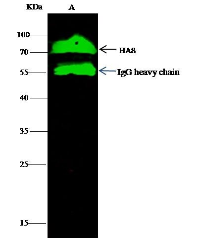
- Human Serum Albumin Immunoprecipitation using: Lane A: 0.5 mg HepG2 Whole Cell Lysate 2 µL with Human Serum Albumin Recombinant Rabbit Monoclonal Antibody (101) (Product # MA5-29022) and 60 μg of Immunomagnetic beads Protein G. Primary antibody: Human Serum Albumin Recombinant Rabbit Monoclonal Antibody (101), at 1:100 dilution. Secondary antibody: Dylight 800-labeled antibody to rabbit IgG (H+L), at 1:5,000 dilution. Developed using the Odyssey technique. Performed under reducing conditions. Predicted band size: 68 kDa. Observed band size: 68 kDa.

Supportive validation
- Submitted by
- Invitrogen Antibodies (provider)
- Main image
- Experimental details
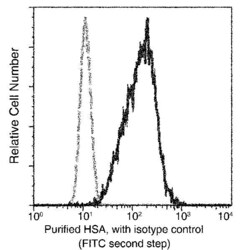
- Flow cytometric analysis of Human HSA expression on HepG2 cells. The cells were treated according to manufacturer’s manual, stained with Human Serum Albumin Recombinant Rabbit Monoclonal Antibody (101) (Product # MA5-29022), then a FITC-conjugated Secondary antibody. The fluorescence histograms were derived from gated events with the forward and side light-scatter characteristics of intact cells.

- Submitted by
- Invitrogen Antibodies (provider)
- Main image

- Experimental details
- Flow cytometric analysis of Human HSA expression on HepG2 cells. The cells were treated according to manufacturer’s manual, stained with Human Serum Albumin Recombinant Rabbit Monoclonal Antibody (101) (Product # MA5-29022), then a FITC-conjugated Secondary antibody. The fluorescence histograms were derived from gated events with the forward and side light-scatter characteristics of intact cells.

Supportive validation
- Submitted by
- Invitrogen Antibodies (provider)
- Main image

- Experimental details
- Human Serum Albumin Immunoprecipitation using: Lane A: 0.5 mg HepG2 Whole Cell Lysate 2 µL with Human Serum Albumin Recombinant Rabbit Monoclonal Antibody (101) (Product # MA5-29022) and 60 μg of Immunomagnetic beads Protein G. Primary antibody: Human Serum Albumin Recombinant Rabbit Monoclonal Antibody (101), at 1:100 dilution. Secondary antibody: Dylight 800-labeled antibody to rabbit IgG (H+L), at 1:5,000 dilution. Developed using the Odyssey technique. Performed under reducing conditions. Predicted band size: 68 kDa. Observed band size: 68 kDa.
- Submitted by
- Invitrogen Antibodies (provider)
- Main image
- Experimental details
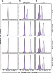
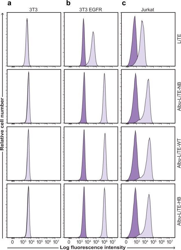
- Fig. 4 Cell target binding properties of Albu-LiTE constructs. Flow cytometry analyses showing target-specific binding with 5 ug ml -1 of the LiTE (102 nM) and Albu-LiTE constructs (46 nM). The LiTE was detected by a C-terminal His-tag directed antibody, while the Albu-LiTE variants were detected using an anti-human serum albumin antibody. a No shift in fluorescence intensity was observed for the double-negative 3T3 cell line, whereas, shifts in fluorescence intensities were seen in b and c . b EGFR positive/CD3 negative 3T3 EGFR cells. c CD3 positive/ EGFR negative Jurkat T-cells. Isotype control antibody was used for background intensity detection.
- Submitted by
- Invitrogen Antibodies (provider)
- Main image
- Experimental details
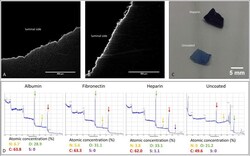
- Figure 2 Coating analyses of exemplary coated BNC grafts: ( A ) Immunofluorescence staining of a cross-sectioned BNC graft against albumin. ( B ) Immunofluorescence staining of a cross-sectioned BNC graft against fibronectin. ( C ) Comparison of toluidine blue staining for heparin-coated and uncoated BNC grafts. ( D ) Results of XPS analyses of the coated BNC grafts. The colored arrows indicate the respective peaks for N (nitrogen), O (oxygen), C (carbon), and S (Sulphur).