Explore
 Validate
 Learn
Learn Western blot
 Immunocytochemistry
Antibody data
- Antibody Data
- Antigen structure
- References [4]
- Comments [0]
- Validations
- Western blot [3]
- Immunohistochemistry [2]
Submit
Validation data
Reference
Comment
Report error
- Product number
- NBP1-32458 - Provider product page

- Provider
- Novus Biologicals
- Proper citation
- Novus Cat#NBP1-32458, RRID:AB_10003946
- Product name
- Rabbit Polyclonal Albumin Antibody
- Antibody type
- Polyclonal
- Description
- Immunogen affinity purified.
- Reactivity
- Human, Mouse, Rat
- Host
- Rabbit
- Isotype
- IgG
- Vial size
- 0.1 ml
- Storage
- Aliquot and store at -20C or -80C. Avoid freeze-thaw cycles.
Submitted references NKG2D and Its Ligand MULT1 Contribute to Disease Progression in a Mouse Model of Multiple Sclerosis.
3-D culture and endothelial cells improve maturity of human pluripotent stem cell-derived hepatocytes.
The in vivo fates of plant viral nanoparticles camouflaged using self-proteins: overcoming immune recognition.
Serum albumin 'camouflage' of plant virus based nanoparticles prevents their antibody recognition and enhances pharmacokinetics.
Legroux L, Moratalla AC, Laurent C, Deblois G, Verstraeten SL, Arbour N
Frontiers in immunology 2019;10:154
Frontiers in immunology 2019;10:154
3-D culture and endothelial cells improve maturity of human pluripotent stem cell-derived hepatocytes.
Ardalani H, Sengupta S, Harms V, Vickerman V, Thomson JA, Murphy WL
Acta biomaterialia 2019 Sep 1;95:371-381
Acta biomaterialia 2019 Sep 1;95:371-381
The in vivo fates of plant viral nanoparticles camouflaged using self-proteins: overcoming immune recognition.
Gulati NM, Pitek AS, Czapar AE, Stewart PL, Steinmetz NF
Journal of materials chemistry. B 2018 Apr 21;6(15):2204-2216
Journal of materials chemistry. B 2018 Apr 21;6(15):2204-2216
Serum albumin 'camouflage' of plant virus based nanoparticles prevents their antibody recognition and enhances pharmacokinetics.
Pitek AS, Jameson SA, Veliz FA, Shukla S, Steinmetz NF
Biomaterials 2016 May;89:89-97
Biomaterials 2016 May;89:89-97
No comments: Submit comment
Supportive validation
- Submitted by
- Novus Biologicals (provider)
- Main image

- Experimental details
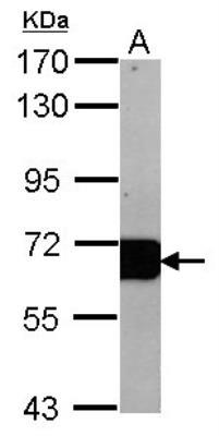
- Western Blot: Albumin Antibody [NBP1-32458] - Sample (20 ug of whole cell lysate) A: mouse Liver 7.5% SDS PAGE diluted at 1:10000
- Submitted by
- Novus Biologicals (provider)
- Main image
- Experimental details
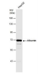
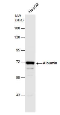
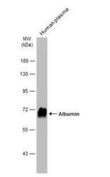
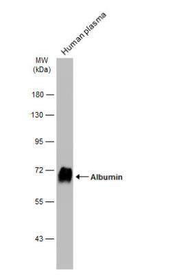
- Western Blot: Albumin Antibody [NBP1-32458] - Whole cell extract (30 ug) was separated by 7.5% SDS-PAGE, and the membrane was blotted with Albumin antibody diluted at 1:1000.
- Submitted by
- Novus Biologicals (provider)
- Main image
- Experimental details
- Western Blot: Albumin Antibody [NBP1-32458] - Human plasma (1 ug) was separated by 7.5% SDS-PAGE, and the membrane was blotted with Albumin antibody diluted at 1:100000. The HRP-conjugated anti-rabbit IgG antibody (NBP2-19301) was used to detect the primary antibody.
Supportive validation
- Submitted by
- Novus Biologicals (provider)
- Main image
- Experimental details
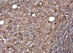
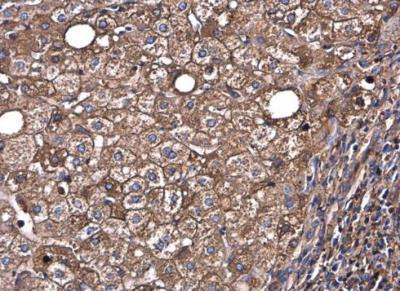
- Immunohistochemistry-Paraffin: Albumin Antibody [NBP1-32458] - Human hepatocellular carcinoma. Albumin antibody diluted at 1:500. Antigen Retrieval: Citrate buffer, pH 6.0, 15 min.
- Submitted by
- Novus Biologicals (provider)
- Main image
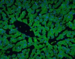
- Experimental details
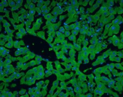
- Immunohistochemistry-Frozen: Albumin Antibody [NBP1-32458] - Albumin antibody detects Albumin protein at cytoplasm in rat liver by immunohistochemical analysis. Sample: Frozen section of rat liver. Albumin antibody diluted at 1:200.