Explore
 Validate
 Learn
LearnMA1-23275
antibody from Invitrogen Antibodies
Targeting: RBBP4
lin-53, NURF55, RbAp48
 Western blot
 Immunocytochemistry Immunoprecipitation
 Immunohistochemistry Chromatin Immunoprecipitation Other assay
Antibody data
- Antibody Data
- Antigen structure
- References [3]
- Comments [0]
- Validations
- Immunocytochemistry [4]
- Immunoprecipitation [1]
- Chromatin Immunoprecipitation [1]
- Other assay [1]
Submit
Validation data
Reference
Comment
Report error
- Product number
- MA1-23275 - Provider product page

- Provider
- Invitrogen Antibodies
- Product name
- RbAp48 Monoclonal Antibody (13D10)
- Antibody type
- Monoclonal
- Antigen
- Other
- Description
- Recommended positive controls: HeLa nuclear extract, SH-SY-5Y, SH-SY-5Y nuclear extract.
- Reactivity
- Human, Mouse, Rat
- Host
- Mouse
- Isotype
- IgG
- Antibody clone number
- 13D10
- Vial size
- 100 μL
- Concentration
- 1 mg/mL
- Storage
- Store at 4°C short term. For long term storage, store at -20°C, avoiding freeze/thaw cycles.
Submitted references Dual retinoblastoma-binding proteins with properties related to a negative regulator of ras in yeast.
Dual retinoblastoma-binding proteins with properties related to a negative regulator of ras in yeast.
A retinoblastoma-binding protein related to a negative regulator of Ras in yeast.
Qian YW, Lee EY
The Journal of biological chemistry 1995 Oct 27;270(43):25507-13
The Journal of biological chemistry 1995 Oct 27;270(43):25507-13
Dual retinoblastoma-binding proteins with properties related to a negative regulator of ras in yeast.
Qian YW, Lee EY
The Journal of biological chemistry 1995 Oct 27;270(43):25507-13
The Journal of biological chemistry 1995 Oct 27;270(43):25507-13
A retinoblastoma-binding protein related to a negative regulator of Ras in yeast.
Qian YW, Wang YC, Hollingsworth RE Jr, Jones D, Ling N, Lee EY
Nature 1993 Aug 12;364(6438):648-52
Nature 1993 Aug 12;364(6438):648-52
No comments: Submit comment
Supportive validation
- Submitted by
- Invitrogen Antibodies (provider)
- Main image

- Experimental details
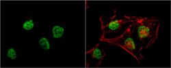
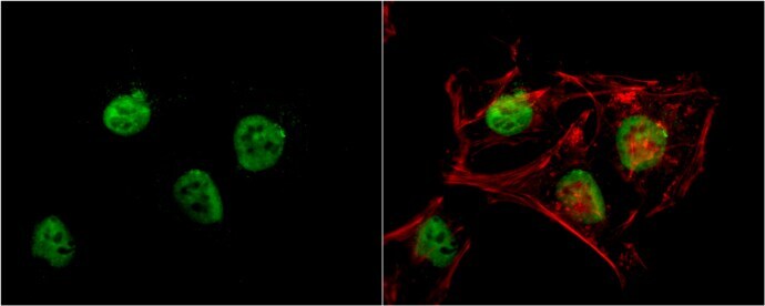
- Immunocytochemistry-Immunofluorescence analysis of RbAp48 was performed in HeLa cells fixed in 4% paraformaldehyde at RT for 15 min. Green: RbAp48 Monoclonal Antibody (13D10) (Product # MA1-23275) diluted at 1:200. Red: phalloidin, a cytoskeleton marker. Blue: Hoechst 33342 staining.

- Submitted by
- Invitrogen Antibodies (provider)
- Main image

- Experimental details
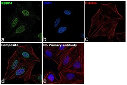
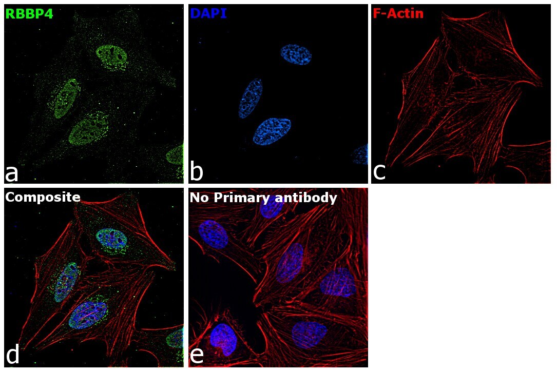
- Immunofluorescence analysis of RbAp48 was performed using 70% confluent log phase HeLa cells. The cells were fixed with 4% paraformaldehyde for 10 minutes, permeabilized with 0.1% Triton™ X-100 for 15 minutes, and blocked with 2% BSA for 1 hour at room temperature. The cells were labeled with RbAp48 Monoclonal Antibody (13D10) (Product # MA1-23275) at 5 µg/mL in 0.1% BSA, incubated at 4 degree Celsius overnight and then labeled with Goat anti-Mouse IgG (H+L) Superclonal™ Recombinant Secondary Antibody, Alexa Fluor® 488 (Product # A28175) at a dilution of 1:2000 for 45 minutes at room temperature (Panel a: green). Nuclei (Panel b: blue) were stained with ProLong™ Diamond Antifade Mountant with DAPI (Product # P36962). F-actin (Panel c: red) was stained with Rhodamine Phalloidin (Product # R415, 1:300). Panel d represents the merged image showing nuclear localization. Panel e represents control cells with no primary antibody to assess background. The images were captured at 60X magnification.

- Submitted by
- Invitrogen Antibodies (provider)
- Main image
- Experimental details
- Immunocytochemistry-Immunofluorescence analysis of RbAp48 was performed in HeLa cells fixed in 4% paraformaldehyde at RT for 15 min. Green: RbAp48 Monoclonal Antibody (13D10) (Product # MA1-23275) diluted at 1:200. Red: phalloidin, a cytoskeleton marker. Blue: Hoechst 33342 staining.
- Submitted by
- Invitrogen Antibodies (provider)
- Main image
- Experimental details
- Immunofluorescence analysis of RbAp48 was performed using 70% confluent log phase HeLa cells. The cells were fixed with 4% paraformaldehyde for 10 minutes, permeabilized with 0.1% Triton™ X-100 for 15 minutes, and blocked with 2% BSA for 1 hour at room temperature. The cells were labeled with RbAp48 Monoclonal Antibody (13D10) (Product # MA1-23275) at 5 µg/mL in 0.1% BSA, incubated at 4 degree Celsius overnight and then labeled with Goat anti-Mouse IgG (H+L) Superclonal™ Recombinant Secondary Antibody, Alexa Fluor® 488 (Product # A28175) at a dilution of 1:2000 for 45 minutes at room temperature (Panel a: green). Nuclei (Panel b: blue) were stained with ProLong™ Diamond Antifade Mountant with DAPI (Product # P36962). F-actin (Panel c: red) was stained with Rhodamine Phalloidin (Product # R415, 1:300). Panel d represents the merged image showing nuclear localization. Panel e represents control cells with no primary antibody to assess background. The images were captured at 60X magnification.
Supportive validation
- Submitted by
- Invitrogen Antibodies (provider)
- Main image

- Experimental details
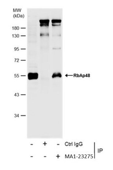
- Immunoprecipitation of RbAp48 was performed in HeLa whole cell extract using 5 µg of RbAp48 Monoclonal Antibody (13D10) (Product # MA1-23275). Samples were transferred to a membrane and probed with RbAp48 Monoclonal Antibody (13D10) as a primary antibody and an HRP-conjugated HRP-conjugated anti mouse IgG was used as a secondary antibody.

Supportive validation
- Submitted by
- Invitrogen Antibodies (provider)
- Main image
- Experimental details
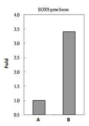
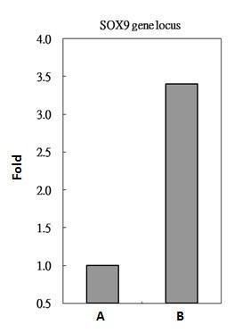
- MA1-23275 antibody immunoprecipitates RbAp48 protein-DNA in ChIP experiments. ChIP Sample: HeLa whole cell lysate/extract with 5 µg preimmune mouse IgG (A) or 5 µg of RbAp48 antibody (Product # MA1-23275) (B). The precipitated DNA was detected by PCR with primer set targeting to SOX9 gene locus.
Supportive validation
- Submitted by
- Invitrogen Antibodies (provider)
- Main image

- Experimental details
- Immunoprecipitation of RbAp48 was performed in HeLa whole cell extract using 5 µg of RbAp48 Monoclonal Antibody (13D10) (Product # MA1-23275). Samples were transferred to a membrane and probed with RbAp48 Monoclonal Antibody (13D10) as a primary antibody and an HRP-conjugated HRP-conjugated anti mouse IgG was used as a secondary antibody.