 Explore
 Validate
 Learn
Learn Western blot
 Immunocytochemistry
Antibody data
- Antibody Data
- Antigen structure
- References [0]
- Comments [0]
- Validations
- Immunocytochemistry [4]
- Immunohistochemistry [1]
- Other assay [1]
Submit
Validation data
Reference
Comment
Report error
- Product number
- PA5-57110 - Provider product page

- Provider
- Invitrogen Antibodies
- Product name
- XRN1 Polyclonal Antibody
- Antibody type
- Polyclonal
- Antigen
- Recombinant protein fragment
- Description
- Immunogen sequence: NVASSVLGKS VFVNWPHLEE ARVVAVSDGE TKFYLEEPPG TQKLYSGRTA PPSKVVHLGD KEQSNWAKEV QGISEHYLRR K Highest antigen sequence identity to the following orthologs: Mouse - 84%, Rat - 85%.
- Reactivity
- Human
- Host
- Rabbit
- Isotype
- IgG
- Vial size
- 100 μL
- Concentration
- 0.4 mg/mL
- Storage
- Store at 4°C short term. For long term storage, store at -20°C, avoiding freeze/thaw cycles.
No comments: Submit comment
Supportive validation
- Submitted by
- Invitrogen Antibodies (provider)
- Main image
- Experimental details
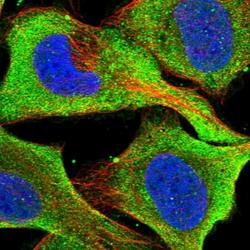
- Immunofluorescent staining of XRN1 in human cell line U-2 OS shows positivity in plasma membrane & cytoplasm. Samples were probed using a XRN1 Polyclonal Antibody (Product # PA5-57110).

- Submitted by
- Invitrogen Antibodies (provider)
- Main image
- Experimental details
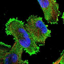
- Immunofluorescent staining of XRN1 in human cell line U-251 MG using a XRN1 Polyclonal Antibody (Product # PA5-57110) shows localization to plasma membrane and cytosol.

- Submitted by
- Invitrogen Antibodies (provider)
- Main image

- Experimental details
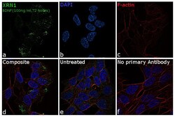
- Immunofluorescence analysis of XRN1 was performed using SH-SY5Y and SH-SY5Y treated with BDNF (100 ng/mL, 72 hours). The cells were fixed with 4% paraformaldehyde for 10 minutes, permeabilized with 0.1% Triton™ X-100 for 10 minutes, and blocked with 1% BSA for 1 hour at room temperature. The cells were labeled with XRN1 Polyclonal Antibody (Product # PA5-57110) at 5 µg/mL concentration in 0.1% BSA and incubated overnight at 4 degree and then labeled with Goat anti-Rabbit IgG (H+L), Superclonal™ Recombinant Secondary Antibody, Alexa Fluor 488 (Product # A27034) at a dilution of 1:2000 for 45 minutes at room temperature (Panel a: green). Nuclei (Panel b: blue) were stained with SlowFade® Gold Antifade Mountant with DAPI (Product # S36938). F-actin (Panel c: red) was stained with Rhodamine Phalloidin (Product # R415, 1:300). Panel d and e represents the merged image showing cytoplasmic localization. Expression of XRN1 was increased upon BDNF treatment in SH-SY5Y cells (Panel d) as compared to untreated SH-SY5Y cells (Panel e). Panel f represents control cells with no primary antibody to assess background. The images were captured at 60X magnification.

- Submitted by
- Invitrogen Antibodies (provider)
- Main image
- Experimental details
- Immunofluorescence analysis of XRN1 was performed using SH-SY5Y and SH-SY5Y treated with BDNF (100 ng/mL, 72 hours). The cells were fixed with 4% paraformaldehyde for 10 minutes, permeabilized with 0.1% Triton™ X-100 for 10 minutes, and blocked with 1% BSA for 1 hour at room temperature. The cells were labeled with XRN1 Polyclonal Antibody (Product # PA5-57110) at 5 µg/mL concentration in 0.1% BSA and incubated overnight at 4 degree and then labeled with Goat anti-Rabbit IgG (Heavy Chain), Superclonal™ Recombinant Secondary Antibody, Alexa Fluor 488 (Product # A27034) at a dilution of 1:2000 for 45 minutes at room temperature (Panel a: green). Nuclei (Panel b: blue) were stained with SlowFade® Gold Antifade Mountant with DAPI (Product # S36938). F-actin (Panel c: red) was stained with Rhodamine Phalloidin (Product # R415, 1:300). Panel d and e represents the merged image showing cytoplasmic localization. Expression of XRN1 was increased upon BDNF treatment in SH-SY5Y cells (Panel d) as compared to untreated SH-SY5Y cells (Panel e). Panel f represents control cells with no primary antibody to assess background. The images were captured at 60X magnification.

Supportive validation
- Submitted by
- Invitrogen Antibodies (provider)
- Main image
- Experimental details
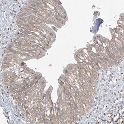
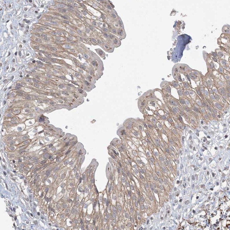
- Immunohistochemical staining of XRN1 in human urinary bladder using a XRN1 Polyclonal Antibody (Product # PA5-57110) shows weak cytoplasmic positivity in urothelial cells.
Supportive validation
- Submitted by
- Invitrogen Antibodies (provider)
- Main image
- Experimental details
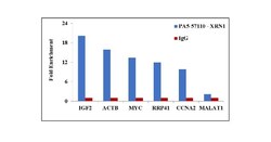
- RNA Immunoprecipitation (RIP) assay of endogenous XRN1 protein using Anti-XRN1 Antibody: RIP assay was performed using Anti-XRN1 Recombinant Rabbit Polyclonal Antibody (Product # PA5-57110, 5 ug) on whole cell lysate from Hep G2 cells exposed to heat shock (45 degrees for 1 hour). Normal Rabbit IgG was used as a negative IP control. RNA purified by RiboPure™ RNA Purification Kit (Product # AM1924) was analyzed by RT-PCR using the Power SYBR® Green RNA-to-CT™ 1-Step Kit (Product # 4389986) with the primers pairs over IGF2, ACTB, MYC, RRP41, CCNA2, IGF2 mRNA and MALAT non-coding RNA. Data is presented as fold enrichment of the antibody signal versus the negative control IgG using the comparative CT method.
