 Explore
 Validate
 Learn
Learn Western blot
Antibody data
- Antibody Data
- Antigen structure
- References [6]
- Comments [0]
- Validations
- Western blot [3]
- Immunoprecipitation [1]
Submit
Validation data
Reference
Comment
Report error
- Product number
- NB500-191 - Provider product page

- Provider
- Novus Biologicals
- Proper citation
- Novus Cat#NB500-191, RRID:AB_2257553
- Product name
- Rabbit Polyclonal Xrn1 Antibody
- Antibody type
- Polyclonal
- Description
- Immunogen affinity purified.
- Reactivity
- Human, Mouse
- Host
- Rabbit
- Isotype
- IgG
- Vial size
- 100 ul
- Concentration
- 1.0 mg/ml
- Storage
- Store at 4C. Do not freeze.
Submitted references A miRNA-responsive cell-free translation system facilitates isolation of hepatitis C virus miRNP complexes.
Chikungunya virus nsP3 blocks stress granule assembly by recruitment of G3BP into cytoplasmic foci.
Hepatitis C virus hijacks P-body and stress granule components around lipid droplets.
Poliovirus-mediated disruption of cytoplasmic processing bodies.
Human Pat1b connects deadenylation with mRNA decapping and controls the assembly of processing bodies.
An RNA pseudoknot is required for production of yellow fever virus subgenomic RNA by the host nuclease XRN1.
Bradrick SS, Nagyal S, Novatt H
RNA (New York, N.Y.) 2013 Aug;19(8):1159-69
RNA (New York, N.Y.) 2013 Aug;19(8):1159-69
Chikungunya virus nsP3 blocks stress granule assembly by recruitment of G3BP into cytoplasmic foci.
Fros JJ, Domeradzka NE, Baggen J, Geertsema C, Flipse J, Vlak JM, Pijlman GP
Journal of virology 2012 Oct;86(19):10873-9
Journal of virology 2012 Oct;86(19):10873-9
Hepatitis C virus hijacks P-body and stress granule components around lipid droplets.
Ariumi Y, Kuroki M, Kushima Y, Osugi K, Hijikata M, Maki M, Ikeda M, Kato N
Journal of virology 2011 Jul;85(14):6882-92
Journal of virology 2011 Jul;85(14):6882-92
Poliovirus-mediated disruption of cytoplasmic processing bodies.
Dougherty JD, White JP, Lloyd RE
Journal of virology 2011 Jan;85(1):64-75
Journal of virology 2011 Jan;85(1):64-75
Human Pat1b connects deadenylation with mRNA decapping and controls the assembly of processing bodies.
Ozgur S, Chekulaeva M, Stoecklin G
Molecular and cellular biology 2010 Sep;30(17):4308-23
Molecular and cellular biology 2010 Sep;30(17):4308-23
An RNA pseudoknot is required for production of yellow fever virus subgenomic RNA by the host nuclease XRN1.
Silva PA, Pereira CF, Dalebout TJ, Spaan WJ, Bredenbeek PJ
Journal of virology 2010 Nov;84(21):11395-406
Journal of virology 2010 Nov;84(21):11395-406
No comments: Submit comment
Supportive validation
- Submitted by
- Novus Biologicals (provider)
- Main image
- Experimental details
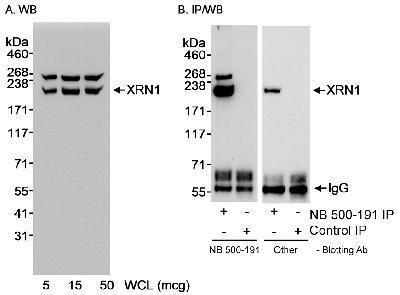
- Western Blot: Xrn1 Antibody [NB500-191] - Detection of Human XRN1 on HeLa whole cell lysate using NB500-191. XRN1 that was immunoprecipitated by NB500-191 was also blotted with a different affinity purified antibody to an upstream region of XRN1.
- Submitted by
- Novus Biologicals (provider)
- Main image
- Experimental details
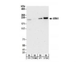
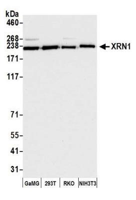
- Western Blot: Xrn1 Antibody [NB500-191] - Whole cell lysate (50 ug) from NIH3T3, TCMK-1, 4T1, and CT26.WT cells. Antibodies: Affinity purified rabbit antiXRN1 antibody used for WB at 0.2 ug/ml. Detection: Chemiluminescence with an exposure time of 3 minutes.
- Submitted by
- Novus Biologicals (provider)
- Main image
- Experimental details
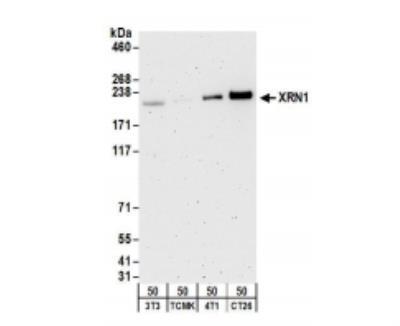
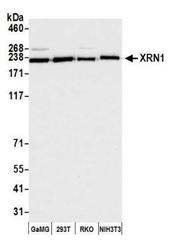
- Western Blot: Xrn1 Antibody [NB500-191] - Detection of human and mouse XRN1 by western blot. Samples:Whole cell lysate (15 ug) from GaMG, HEK293T, RKO, and NIH 3T3 cells prepared using NETN lysis buffer. Antibody: Affinity purified rabbit anti-XRN1 antibody NB500-191 used for WB at 0.04 ug/ml. Detection: Chemiluminescence with an exposure time of 10 seconds.
Supportive validation
- Submitted by
- Novus Biologicals (provider)
- Main image
- Experimental details
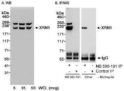
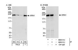
- Immunoprecipitation: Xrn1 Antibody [NB500-191] - Detection of human XRN1 by western blot and immunoprecipitation. Samples: Whole cell lysate from HeLa (5, 15 and 50 ug for WB; 1 mg for IP, 20% of IP loaded) and HEK293T (T; 50 ug) cells. Antibodies: Affinity purified rabbit anti-XRN1 antibody NB500-191 (lot 3) used for WB at 0.1 ug/ml (A) and 1 ug/ml (B) and used for IP at 3 ug/mg lysate. XRN1 was also immunoprecipitated by a previous lot of this antibody (lot 2). Detection: Chemiluminescence with exposure times of 30 seconds (A) and 10 seconds (B).
