 Explore
 Validate
 Learn
Learn Western blot
Antibody data
- Antibody Data
- Antigen structure
- References [0]
- Comments [0]
- Validations
- Western blot [1]
- Immunocytochemistry [1]
- Immunohistochemistry [1]
Submit
Validation data
Reference
Comment
Report error
- Product number
- TA328699 - Provider product page

- Provider
- OriGene
- Product name
- Rabbit Polyclonal Anti-GABA (A) alpha3 Receptor (extracellular)
- Antibody type
- Polyclonal
- Description
- Rabbit Polyclonal Anti-GABA (A) alpha3 Receptor (extracellular)
- Host
- Rabbit
- Conjugate
- Unconjugated
- Epitope
- GABRA3
- Antibody clone number
- NULL
- Vial size
- 200 µl
- Concentration
- NULL
No comments: Submit comment
Supportive validation
- Submitted by
- OriGene (provider)
- Main image
- Experimental details
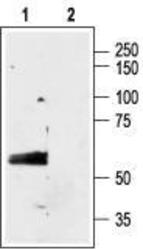
- Western blot analysis of rat brain membranes: 1. Anti-GABA(A) a3 Receptor (extracellular) antibody, (1:200). 2. Anti-GABA(A) a3 Receptor (extracellular) antibody preincubated with the control peptide antigen.
- Validation comment
- WB

Supportive validation
- Submitted by
- OriGene (provider)
- Main image
- Experimental details
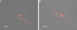
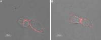
- Expression of GABA(A) a3 Receptor in rat insulinoma cell line. Immunocytochemical staining of intact living rat RIN-m cells using Anti- GABA(A) a3 Receptor (extracellular) antibody, (1:50) followed by goat anti-rabbit-AlexaFluor-550 secondary antibody. Extracellular staining (red) merged with live view of the cells.
- Validation comment
- IF
Supportive validation
- Submitted by
- OriGene (provider)
- Main image
- Experimental details
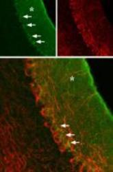
- Expression of GABA(A) a3 Receptor in rat cerebellum. Immunohistochemical staining of rat cerebellum using Anti-GABA(A) a3 Receptor (extracellular), (green), (1:100). GABA(A) a3 Receptor is localized to the molecular layer (asterisk) and a portion of the Purkinje cell body (arrows), which is outlined by axonal staining using mouse anti-neurofilament 200 (red).
- Validation comment
- IHC
