Explore
 Validate
 Learn
Learn Western blot
Antibody data
- Antibody Data
- Antigen structure
- References [0]
- Comments [0]
- Validations
- Western blot [1]
- Immunocytochemistry [1]
- Immunohistochemistry [1]
Submit
Validation data
Reference
Comment
Report error
- Product number
- AGA-003-25UL - Provider product page

- Provider
- Invitrogen Antibodies
- Product name
- GABA(A) alpha 3 Receptor (extracellular) Polyclonal Antibody
- Antibody type
- Polyclonal
- Antigen
- Other
- Reactivity
- Human, Mouse, Rat
- Host
- Rabbit
- Isotype
- IgG
- Vial size
- 25 µL
- Concentration
- 0.75 mg/mL
- Storage
- -20° C, Avoid Freeze/Thaw Cycles
No comments: Submit comment
Supportive validation
- Submitted by
- Invitrogen Antibodies (provider)
- Main image
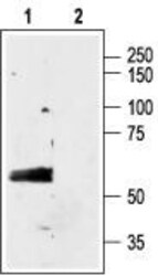
- Experimental details
- Western blot analysis of rat brain membranes: - 1. Anti-GABA (A) alpha 3 Receptor (extracellular) Antibody (#AGA-003), (1:200). 2. Anti-GABA (A) alpha 3 Receptor (extracellular) Antibody preincubated with GABA (A) alpha 3 Receptor (extracellular) Blocking Peptide (#BLP-GA003).
Supportive validation
- Submitted by
- Invitrogen Antibodies (provider)
- Main image
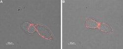
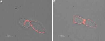
- Experimental details
- Expression of GABRA3 in rat insulinoma cell line - Cell surface detection of GABRA3 in intact living rat RIN-m cells using Anti-GABA (A) alpha 3 Receptor (extracellular) Antibody (#AGA-003), (1:50) followed by goat Anti-rabbit-AlexaFluor-550 secondary Antibody . Extracellular staining (red) merged with live view of the cells.
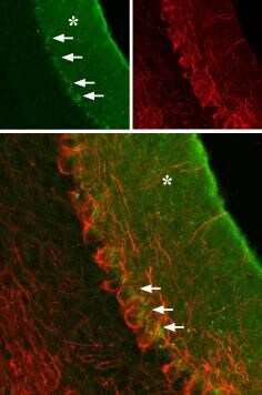
Supportive validation
- Submitted by
- Invitrogen Antibodies (provider)
- Main image
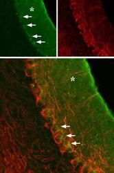
- Experimental details
- Expression ofGABRA3 in rat cerebellum - Immunohistochemical staining of rat cerebellum using Anti-GABA (A) alpha 3 Receptor (extracellular) Antibody (#AGA-003), (green), (1:100). GABRA3 is localized to the molecular layer (asterisk) and a portion of the Purkinje cell body (arrows), which is outlined by axonal staining using mouse Anti-neurofilament 200 (red).