Explore
 Validate
 Learn
Learn Western blot
 Flow cytometry
Antibody data
- Antibody Data
- Antigen structure
- References [5]
- Comments [0]
- Validations
- Flow cytometry [2]
- Other assay [3]
Submit
Validation data
Reference
Comment
Report error
- Product number
- 14-9230-82 - Provider product page

- Provider
- Invitrogen Antibodies
- Product name
- CD230 (PrP) Monoclonal Antibody (4D5), eBioscience™
- Antibody type
- Monoclonal
- Antigen
- Other
- Description
- Description: CD230 (prion protein; PrP^C) is a 35 kD glycoprotein, attached to the cell surface via a GPI-anchor. While the function of CD230 is unclear, prion protein has been the focus of research based on its involvement in transmissible spongiform encephalopathies (TSE), including Creutzfeld-Jakob disease in humans. CD230 undergoes a conformational change in which the cellular PrP^C form is converted to the scrapie PrP^Sc form. This conversion is essential for the infectiousness of prion-based diseases. CD230 is predominantly expressed on the surface of lymphocytes and monocytes, with weak expression on granulocytes and erythrocytes. In platelets, CD230 is expressed primarily intracellularly, and is upregulated to the surface upon activation. Applications Reported: This 4D5 antibody has been reported for use in flow cytometric analysis and immunoprecipitation. The 4D5 antibody has also been found useful for immunoblotting, recognizing a protein of approximately 35 kD under non-reducing conditions. Applications Tested: The 4D5 antibody has been tested by flow cytometric analysis of normal human peripheral blood cells. This can be used at less than or equal to 0.5 µg per test. A test is defined as the amount (µg) of antibody that will stain a cell sample in a final volume of 100 µL. Cell number should be determined empirically but can range from 10^5 to 10^8 cells/test. It is recommended that the antibody be carefully titrated for optimal performance in the assay of interest. Purity: Greater than 90%, as determined by SDS-PAGE. Aggregation: Less than 10%, as determined by HPLC. Filtration: 0.2 µm post-manufacturing filtered.
- Reactivity
- Human
- Host
- Mouse
- Isotype
- IgG
- Antibody clone number
- 4D5
- Vial size
- 100 μg
- Concentration
- 0.5 mg/mL
- Storage
- 4°C
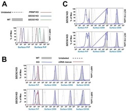
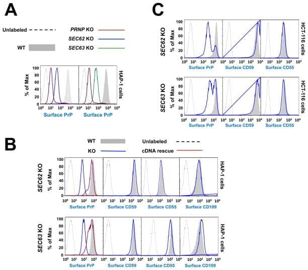
Submitted references A knockout cell library of GPI biosynthetic genes for functional studies of GPI-anchored proteins.
Comparative Haploid Genetic Screens Reveal Divergent Pathways in the Biogenesis and Trafficking of Glycophosphatidylinositol-Anchored Proteins.
Activated platelets of patients with paroxysmal nocturnal hemoglobinuria express cellular prion protein.
Differential constitutive and activation-dependent expression of prion protein in human peripheral blood leucocytes.
Distribution of cell-associated prion protein in normal adult blood determined by flow cytometry.
Liu SS, Liu YS, Guo XY, Murakami Y, Yang G, Gao XD, Kinoshita T, Fujita M
Communications biology 2021 Jun 23;4(1):777
Communications biology 2021 Jun 23;4(1):777
Comparative Haploid Genetic Screens Reveal Divergent Pathways in the Biogenesis and Trafficking of Glycophosphatidylinositol-Anchored Proteins.
Davis EM, Kim J, Menasche BL, Sheppard J, Liu X, Tan AC, Shen J
Cell reports 2015 Jun 23;11(11):1727-36
Cell reports 2015 Jun 23;11(11):1727-36
Activated platelets of patients with paroxysmal nocturnal hemoglobinuria express cellular prion protein.
Holada K, Simak J, Risitano AM, Maciejewski J, Young NS, Vostal JG
Blood 2002 Jul 1;100(1):341-3
Blood 2002 Jul 1;100(1):341-3
Differential constitutive and activation-dependent expression of prion protein in human peripheral blood leucocytes.
Dürig J, Giese A, Schulz-Schaeffer W, Rosenthal C, Schmücker U, Bieschke J, Dührsen U, Kretzschmar HA
British journal of haematology 2000 Mar;108(3):488-95
British journal of haematology 2000 Mar;108(3):488-95
Distribution of cell-associated prion protein in normal adult blood determined by flow cytometry.
Barclay GR, Hope J, Birkett CR, Turner ML
British journal of haematology 1999 Dec;107(4):804-14
British journal of haematology 1999 Dec;107(4):804-14
No comments: Submit comment
Supportive validation
- Submitted by
- Invitrogen Antibodies (provider)
- Main image

- Experimental details
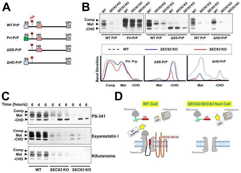
- Staining of normal human peripheral blood cells with 0.25 µg of Purified Mouse IgG1 kappa Isotype Control (Product # 14-4714-82) (open histogram) or 0.25 µg of Anti-Human CD230 (PrP) Purified (filled histogram) followed by F (ab')2 Anti-Mouse IgG PE (Product # 12-4012). Cells in the lymphocyte gate were used for analysis.

- Submitted by
- Invitrogen Antibodies (provider)
- Main image
- Experimental details
- Staining of normal human peripheral blood cells with 0.25 µg of Purified Mouse IgG1 kappa Isotype Control (Product # 14-4714-82) (open histogram) or 0.25 µg of Anti-Human CD230 (PrP) Purified (filled histogram) followed by F (ab')2 Anti-Mouse IgG PE (Product # 12-4012). Cells in the lymphocyte gate were used for analysis.

Supportive validation
- Submitted by
- Invitrogen Antibodies (provider)
- Main image
- Experimental details
- NULL
- Submitted by
- Invitrogen Antibodies (provider)
- Main image
- Experimental details
- NULL
- Submitted by
- Invitrogen Antibodies (provider)
- Main image
- Experimental details
- NULL