Explore
 Validate
 Learn
Learn Western blot
 Immunocytochemistry
Antibody data
- Antibody Data
- Antigen structure
- References [2]
- Comments [0]
- Validations
- Immunocytochemistry [3]
- Immunohistochemistry [2]
- Flow cytometry [1]
- Other assay [1]
Submit
Validation data
Reference
Comment
Report error
- Product number
- MA5-32202 - Provider product page

- Provider
- Invitrogen Antibodies
- Product name
- CD230 (PrP) Recombinant Rabbit Monoclonal Antibody (SC57-05)
- Antibody type
- Monoclonal
- Antigen
- Synthetic peptide
- Description
- Recombinant rabbit monoclonal antibodies are produced using in vitro expression systems. The expression systems are developed by cloning in the specific antibody DNA sequences from immunoreactive rabbits. Then, individual clones are screened to select the best candidates for production. The advantages of using recombinant rabbit monoclonal antibodies include: better specificity and sensitivity, lot-to-lot consistency, animal origin-free formulations, and broader immunoreactivity to diverse targets due to larger rabbit immune repertoire.
- Reactivity
- Human, Mouse, Rat
- Host
- Rabbit
- Isotype
- IgG
- Antibody clone number
- SC57-05
- Vial size
- 100 μL
- Concentration
- 1 mg/mL
- Storage
- Store at 4°C short term. For long term storage, store at -20°C, avoiding freeze/thaw cycles.
Submitted references Extracellular Protein Aggregates Colocalization and Neuronal Dystrophy in Comorbid Alzheimer's and Creutzfeldt-Jakob Disease: A Micromorphological Pilot Study on 20 Brains.
Extracellular Prion Protein Aggregates in Nine Gerstmann-Sträussler-Scheinker Syndrome Subjects with Mutation P102L: A Micromorphological Study and Comparison with Literature Data.
Jankovska N, Olejar T, Matej R
International journal of molecular sciences 2021 Feb 20;22(4)
International journal of molecular sciences 2021 Feb 20;22(4)
Extracellular Prion Protein Aggregates in Nine Gerstmann-Sträussler-Scheinker Syndrome Subjects with Mutation P102L: A Micromorphological Study and Comparison with Literature Data.
Jankovska N, Matej R, Olejar T
International journal of molecular sciences 2021 Dec 10;22(24)
International journal of molecular sciences 2021 Dec 10;22(24)
No comments: Submit comment
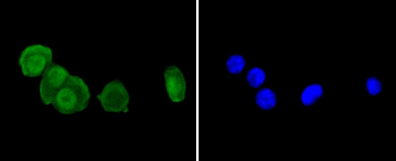
Supportive validation
- Submitted by
- Invitrogen Antibodies (provider)
- Main image
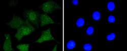
- Experimental details
- Immunocytochemical analysis of CD230 (PrP) in SHG-44 cells using a CD230 (PrP) Monoclonal antibody (Product # MA5-32202) as seen in green. The nuclear counter stain is DAPI (blue). Cells were fixed in paraformaldehyde, permeabilised with 0.25% Triton X100/PBS.
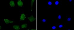
- Submitted by
- Invitrogen Antibodies (provider)
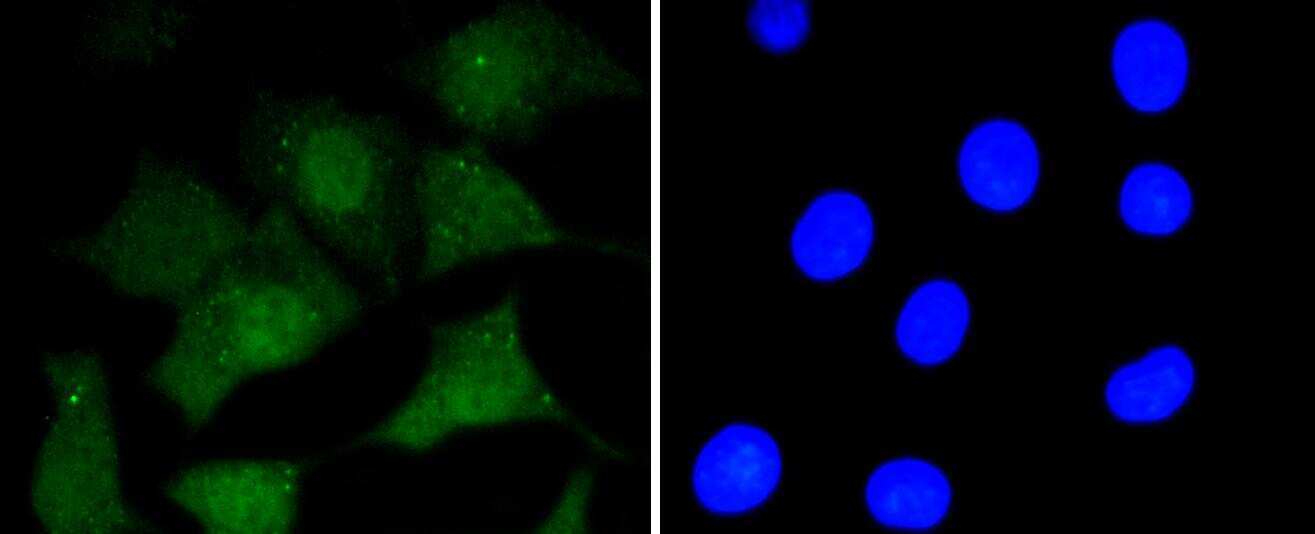
- Main image
- Experimental details
- Immunocytochemical analysis of CD230 (PrP) in N2A cells using a CD230 (PrP) Monoclonal antibody (Product # MA5-32202) as seen in green. The nuclear counter stain is DAPI (blue). Cells were fixed in paraformaldehyde, permeabilised with 0.25% Triton X100/PBS.
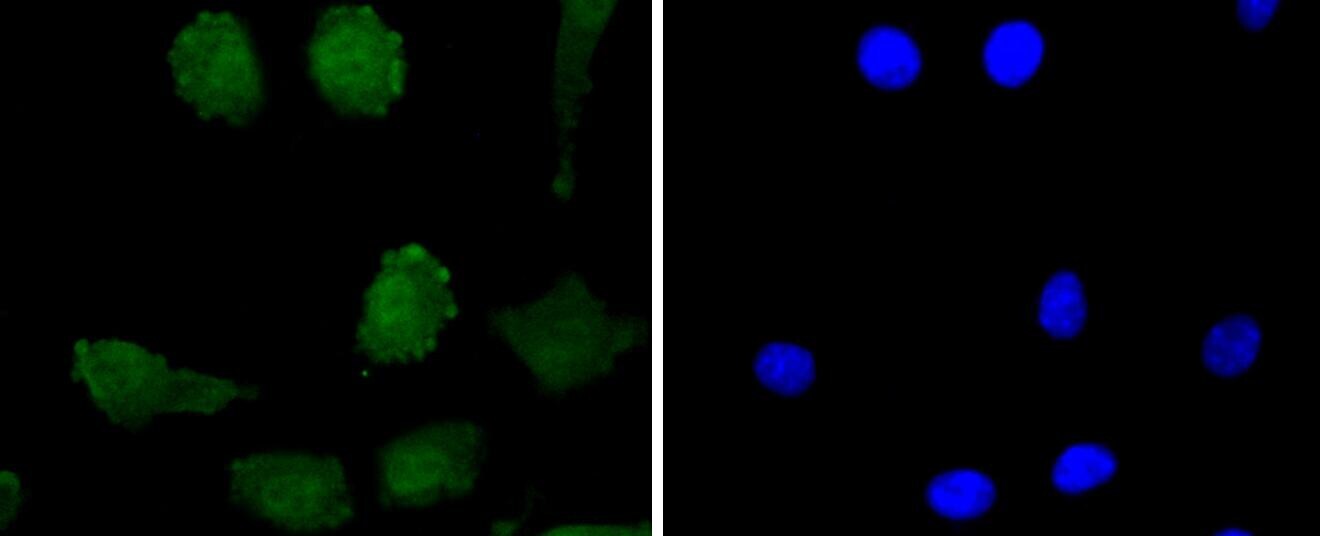
- Submitted by
- Invitrogen Antibodies (provider)
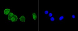
- Main image
- Experimental details
- Immunocytochemical analysis of CD230 (PrP) in SH-SY-5Y cells using a CD230 (PrP) Monoclonal antibody (Product # MA5-32202) as seen in green. The nuclear counter stain is DAPI (blue). Cells were fixed in paraformaldehyde, permeabilised with 0.25% Triton X100/PBS.
Supportive validation
- Submitted by
- Invitrogen Antibodies (provider)
- Main image
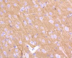
- Experimental details
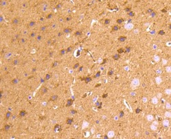
- Immunohistochemical analysis of CD230 (PrP) of paraffin-embedded rat brain tissue using a CD230-PrP Monoclonal antibody (Product #MA5-32202). Counter stained with hematoxylin.

- Submitted by
- Invitrogen Antibodies (provider)
- Main image
- Experimental details
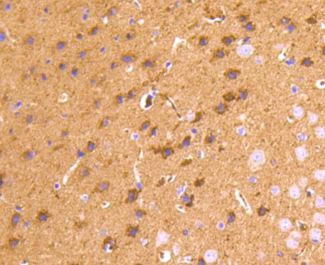
- Immunohistochemical analysis of CD230 (PrP) of paraffin-embedded Mouse brain tissue using a CD230-PrP Monoclonal antibody (Product #MA5-32202). Counter stained with hematoxylin.
Supportive validation
- Submitted by
- Invitrogen Antibodies (provider)
- Main image
- Experimental details
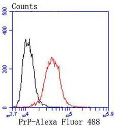
- Flow Cytometric analysis of CD230 (PrP) in SH-SY-5Y cells using a CD230 (PrP) Monoclonal Antibody (Product # MA5-32202) at a dilution of 1:50, as seen in red compared with an unlabelled control (cells without incubation with primary antibody; black). Alexa Fluor 488-conjugated goat anti rabbit IgG was used as the secondary antibody.
Supportive validation
- Submitted by
- Invitrogen Antibodies (provider)
- Main image

- Experimental details
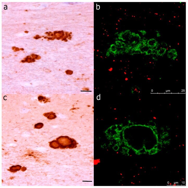
- Figure 1 Illustration--Parallel observation of ( a , b ) multicentric and ( c , d ) solitary kuru-like plaques with centrally bright cores visualized using immunohistochemistry and immunofluorescence. ( a , c ) Primary antibody in immunohistochemical images: PrP (mouse monoclonal antibody). The secondary antibody was conjugated with horseradish peroxidase staining DAB. The original magnification was 100x. ( b , d ) Primary antibodies in immunofluorescent images: PrP (rabbit recombinant monoclonal antibody, green color) + AT8 (mouse monoclonal antibody, red color). The secondary antibody was conjugated with either Alexa Fluor (r) 488 (anti-rabbit IgG, green) or Alexa Fluor (r) 568 (anti-mouse IgG, red). Scale bars indicate 25 um in ( a , b ) and 10 um in ( c , d ).