Explore
 Validate
 Learn
Learn Western blot
 ELISA
ELISAAntibody data
- Antibody Data
- Antigen structure
- References [5]
- Comments [0]
- Validations
- Western blot [1]
- Immunocytochemistry [1]
Submit
Validation data
Reference
Comment
Report error
- Product number
- MA1-750 - Provider product page

- Provider
- Invitrogen Antibodies
- Product name
- PrP Monoclonal Antibody (F89/160.1.5)
- Antibody type
- Monoclonal
- Antigen
- Synthetic peptide
- Description
- MA1-750 detects prion protein (PrP) protein from a variety of species that have the conserved n-IHFG-c epitope, including agriculturally important animal species such as sheep, bovine, deer, and elk. MA1-750 has been successfully used in Western blot, immunohistochemistry and ELISA procedures. By Western blot, this antibody detects a 33-35 kDa protein from normal animals and a 27-30 kDa protein which represents PrP in brain protease treated tissue extracts from infected animals. Immunohistochemical staining of PrP(Sc) in infected sheep brain with MA1-750 results in intense staining of the spongiform lesions. In immunohistochemical procedures this antibody detects only the PrP(Sc) and not PrP(C). The MA1-750 immunogen is a synthetic peptide corresponding to residues S(146) R P L I H F G S D Y E D R(159) of bovine PrP. This peptide (Cat. # PEP-051) is available for use in neutralization and control experiments. MA1-750 is known to specifically recognize a conserved epitope of the PrP(Sc) protein comprising the amino acids n-IHFG-c.
- Reactivity
- Human, Mouse, Bovine
- Host
- Mouse
- Isotype
- IgG
- Antibody clone number
- F89/160.1.5
- Vial size
- 200 µg
- Concentration
- 1 mg/mL
- Storage
- -20° C, Avoid Freeze/Thaw Cycles
Submitted references Differentiating blood samples from scrapie infected and non-infected hamsters by detecting disease-associated prion proteins using Multimer Detection System.
Preclinical diagnosis of scrapie by immunohistochemistry of third eyelid lymphoid tissue.
The pattern of prion-related protein expression in the gastrointestinal tract.
Preclinical detection of PrPSc in nictitating membrane lymphoid tissue of sheep.
Monoclonal antibody F89/160.1.5 defines a conserved epitope on the ruminant prion protein.
An SS, Lim KT, Oh HJ, Lee BS, Zukic E, Ju YR, Yokoyama T, Kim SY, Welker E
Biochemical and biophysical research communications 2010 Feb 19;392(4):505-9
Biochemical and biophysical research communications 2010 Feb 19;392(4):505-9
Preclinical diagnosis of scrapie by immunohistochemistry of third eyelid lymphoid tissue.
O'Rourke KI, Baszler TV, Besser TE, Miller JM, Cutlip RC, Wells GA, Ryder SJ, Parish SM, Hamir AN, Cockett NE, Jenny A, Knowles DP
Journal of clinical microbiology 2000 Sep;38(9):3254-9
Journal of clinical microbiology 2000 Sep;38(9):3254-9
The pattern of prion-related protein expression in the gastrointestinal tract.
Pammer J, Cross HS, Frobert Y, Tschachler E, Oberhuber G
Virchows Archiv : an international journal of pathology 2000 May;436(5):466-72
Virchows Archiv : an international journal of pathology 2000 May;436(5):466-72
Preclinical detection of PrPSc in nictitating membrane lymphoid tissue of sheep.
O'Rourke KI, Baszler TV, Parish SM, Knowles DP
The Veterinary record 1998 May 2;142(18):489-91
The Veterinary record 1998 May 2;142(18):489-91
Monoclonal antibody F89/160.1.5 defines a conserved epitope on the ruminant prion protein.
O'Rourke KI, Baszler TV, Miller JM, Spraker TR, Sadler-Riggleman I, Knowles DP
Journal of clinical microbiology 1998 Jun;36(6):1750-5
Journal of clinical microbiology 1998 Jun;36(6):1750-5
No comments: Submit comment
Supportive validation
- Submitted by
- Invitrogen Antibodies (provider)
- Main image

- Experimental details
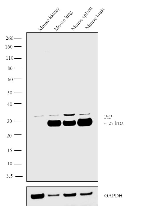
- Western blot analysis was performed on tissue extracts (30 µg lysate) of Mouse kidney (Lane 1), Mouse lung (Lane 2), Mouse spleen (Lane 3) and Mouse brain (Lane 4). The blot was probed with Anti-PrP Monoclonal Antibody (Product # MA1-750, 1:1000 dilution) and detected by chemiluminescence using Goat anti-Mouse IgG (H+L) Superclonal™ Secondary Antibody, HRP conjugate (Product # A28177, 0.25 µg/ml, 1:4000 dilution). A 27 kDa band corresponding to PrP was observed across the tissues positive for PrP, while this band was absent in Mouse kidney tissue lysate which is reported to be negative.
Supportive validation
- Submitted by
- Invitrogen Antibodies (provider)
- Main image

- Experimental details
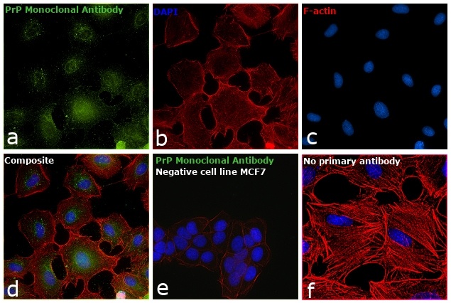
- Immunofluorescence analysis of PrP was performed using 70% confluent log phase A549 or MCF7 cells. The cells were fixed with 4% paraformaldehyde for 10 minutes, permeabilized with 0.1% Triton™ X-100 for 15 minutes, and blocked with 1% BSA for 1 hour at room temperature. The cells were labeled with Anti-PrP Monoclonal Antibody (F89/160.1.5) (Product # MA1-750) at 1:200 dilution in 0.1% BSA, incubated at 4 degree Celsius overnight and then labeled with Goat anti-Mouse IgG (H+L) Superclonal™ Secondary Antibody, Alexa Fluor® 488 conjugate (Product # A28175) at a dilution of 1:2000 for 45 minutes at room temperature (Panel a: green).Nuclei (Panel b: blue) were stained with ProLong™ Diamond Antifade Mountant with DAPI (Product # P36962). F-actin (Panel c: red) was stained with Rhodamine Phalloidin (Product # R415, 1:300). Panel d represents the merged image showing cytosolic localization. Panel e shows no staining for PrP in MCF7 which is a negative model. Panel f represents control A549 cells with no primary antibody to assess background. The images were captured at 60X magnification.