Explore
 Validate
 Learn
Learn Immunohistochemistry
Antibody data
- Antibody Data
- Antigen structure
- References [1]
- Comments [0]
- Validations
- Immunohistochemistry [1]
Submit
Validation data
Reference
Comment
Report error
- Product number
- MAB5140 - Provider product page

- Provider
- R&D Systems
- Product name
- Human SMOC-2 Antibody
- Antibody type
- Monoclonal
- Description
- Protein A or G purified from hybridoma culture supernatant. Detects human SMOC-2 in direct ELISAs and Western blots. In Western blots, approximately 15% cross-reactivity with recombinant human (rh) SMOC-1 and no cross-reactivity with recombinant mouse SMOC-2 is observed.
- Reactivity
- Human
- Host
- Mouse
- Conjugate
- Unconjugated
- Antigen sequence
Q9H3U7- Isotype
- IgG
- Antibody clone number
- 667713
- Vial size
- 100 ug
- Concentration
- LYOPH
- Storage
- Use a manual defrost freezer and avoid repeated freeze-thaw cycles. 12 months from date of receipt, -20 to -70 °C as supplied. 1 month, 2 to 8 °C under sterile conditions after reconstitution. 6 months, -20 to -70 °C under sterile conditions after reconstitution.
Submitted references Silencing SMOC2 ameliorates kidney fibrosis by inhibiting fibroblast to myofibroblast transformation.
Gerarduzzi C, Kumar RK, Trivedi P, Ajay AK, Iyer A, Boswell S, Hutchinson JN, Waikar SS, Vaidya VS
JCI insight 2017 Apr 20;2(8)
JCI insight 2017 Apr 20;2(8)
No comments: Submit comment
Supportive validation
- Submitted by
- R&D Systems (provider)
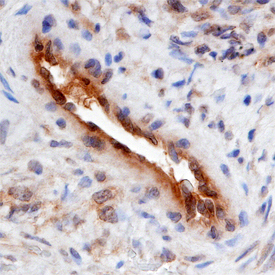
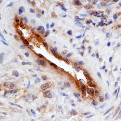
- Main image
- Experimental details
- SMOC-2 in Human Ovarian Cancer Tissue. SMOC-2 was detected in immersion fixed paraffin-embedded sections of human ovarian cancer tissue using Human SMOC-2 Monoclonal Antibody (Catalog # MAB5140) at 15 µg/mL overnight at 4 °C. Before incubation with the primary antibody, tissue was subjected to heat-induced epitope retrieval using Antigen Retrieval Reagent-Basic (Catalog # CTS013). Tissue was stained using the Anti-Mouse HRP-DAB Cell & Tissue Staining Kit (brown; Catalog # CTS002) and counterstained with hematoxylin (blue). Specific staining was localized to endothelial cells. View our protocol for Chromogenic IHC Staining of Paraffin-embedded Tissue Sections.