 Explore
 Validate
 Learn
Learn Western blot
 Flow cytometry
Antibody data
- Antibody Data
- Antigen structure
- References [2]
- Comments [0]
- Validations
- Flow cytometry [1]
- Other assay [1]
Submit
Validation data
Reference
Comment
Report error
- Product number
- PA5-80123 - Provider product page

- Provider
- Invitrogen Antibodies
- Product name
- Thrombospondin 2 Polyclonal Antibody
- Antibody type
- Polyclonal
- Antigen
- Synthetic peptide
- Description
- Reconstitute with 0.2 mL of distilled water to yield a concentration of 500 µg/mL. Positive Control - WB: human Hela whole cell, human MCF-7 whole cell, human U87 whole cell, human U251 whole cell.
- Reactivity
- Human
- Host
- Rabbit
- Isotype
- IgG
- Vial size
- 100 μg
- Concentration
- 500 μg/mL
- Storage
- -20°C
Submitted references Tsp2 Facilitates Tumor-associated Fibroblasts Formation and Promotes Tumor Progression in Retroperitoneal Liposarcoma.
MicroRNA‑203a‑3p is a candidate tumor suppressor that targets thrombospondin 2 in colorectal carcinoma.
Xu C, Yan L, Guan X, Wang Z, Wu J, Lv A, Liu D, Liu F, Dong B, Zhao M, Jia L, Tian X, Hao C
International journal of biological sciences 2022;18(13):5038-5055
International journal of biological sciences 2022;18(13):5038-5055
MicroRNA‑203a‑3p is a candidate tumor suppressor that targets thrombospondin 2 in colorectal carcinoma.
Qian Z, Gong L, Mou Y, Han Y, Zheng S
Oncology reports 2019 Nov;42(5):1825-1832
Oncology reports 2019 Nov;42(5):1825-1832
No comments: Submit comment
Supportive validation
- Submitted by
- Invitrogen Antibodies (provider)
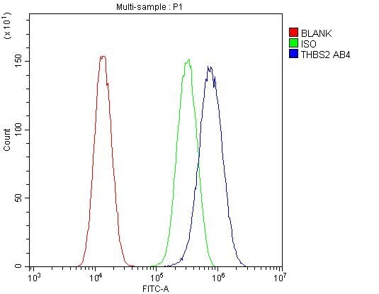
- Main image

- Experimental details
- Flow Cytometry of Thrombospondin 2 in U-87MG cells (blue line), isotype control rabbit IgG (green line) and unlabeled (red line). Samples were blocked with 10% goat serum, incubated with Thrombospondin 2 Polyclonal Antibody (Product # PA5-80123) at a dilution of 1 μg (per 1x10^6 cells), followed by DyLight®488 conjugated goat anti-rabbit IgG (for 30 minutes at 20°C) using 5-10 μg (per 1x10^6 cells) dilution.
Supportive validation
- Submitted by
- Invitrogen Antibodies (provider)
- Main image
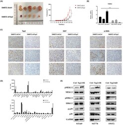
- Experimental details
- Tsp2 downregulation suppressed xenograft tumor growth in NOD/SCID mice and the potential mechanism was elucidated by protein microarray. (A) On day 60, the volume of xenograft tumors formed by SW872 shTsp2 cells were smaller than tumors formed by SW872 shCtrl cells (p
