Explore
 Validate
 Learn
Learn Western blot
 Immunocytochemistry
Antibody data
- Antibody Data
- Antigen structure
- References [2]
- Comments [0]
- Validations
- Immunocytochemistry [2]
- Immunohistochemistry [3]
- Other assay [2]
Submit
Validation data
Reference
Comment
Report error
- Product number
- PA5-30678 - Provider product page

- Provider
- Invitrogen Antibodies
- Product name
- PHF10 Polyclonal Antibody
- Antibody type
- Polyclonal
- Antigen
- Recombinant full-length protein
- Description
- Recommended positive controls: HeLa, HeLa nuclear extract, mouse colon. Predicted reactivity: Mouse (93%), Rat (94%), Bovine (80%). Store product as a concentrated solution. Centrifuge briefly prior to opening the vial.
- Reactivity
- Human, Mouse, Rat
- Host
- Rabbit
- Isotype
- IgG
- Vial size
- 100 μL
- Concentration
- 1.06 mg/mL
- Storage
- Store at 4°C short term. For long term storage, store at -20°C, avoiding freeze/thaw cycles.
Submitted references Gut Bacteria Selectively Altered by Sennoside A Alleviate Type 2 Diabetes and Obesity Traits.
A Genome-wide CRISPR Screen Reveals a Role for the Non-canonical Nucleosome-Remodeling BAF Complex in Foxp3 Expression and Regulatory T Cell Function.
Wei Z, Shen P, Cheng P, Lu Y, Wang A, Sun Z
Oxidative medicine and cellular longevity 2020;2020:2375676
Oxidative medicine and cellular longevity 2020;2020:2375676
A Genome-wide CRISPR Screen Reveals a Role for the Non-canonical Nucleosome-Remodeling BAF Complex in Foxp3 Expression and Regulatory T Cell Function.
Loo CS, Gatchalian J, Liang Y, Leblanc M, Xie M, Ho J, Venkatraghavan B, Hargreaves DC, Zheng Y
Immunity 2020 Jul 14;53(1):143-157.e8
Immunity 2020 Jul 14;53(1):143-157.e8
No comments: Submit comment
Supportive validation
- Submitted by
- Invitrogen Antibodies (provider)
- Main image

- Experimental details
- Immunocytochemistry-Immunofluorescence analysis of PHF10 was performed in HeLa cells fixed in 4% paraformaldehyde at RT for 15 min. Green: PHF10 Polyclonal Antibody (Product # PA5-30678) diluted at 1:500. Red: Phalloidin, a cytoskeleton marker. Blue: Hoechst 33342 staining.

Supportive validation
- Submitted by
- Invitrogen Antibodies (provider)
- Main image

- Experimental details
- Immunohistochemistry (Paraffin) analysis of PHF10 was performed in paraffin-embedded mouse lung tissue using PHF10 Polyclonal Antibody (Product # PA5-30678) at a dilution of 1:500.
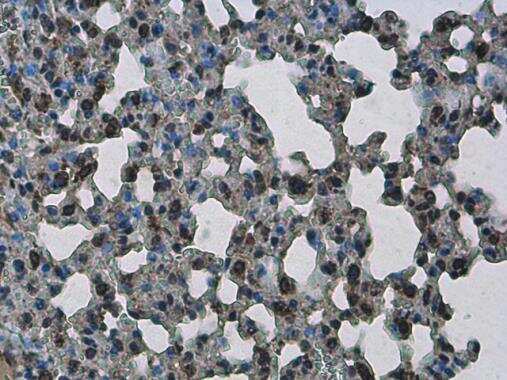
- Submitted by
- Invitrogen Antibodies (provider)
- Main image
- Experimental details
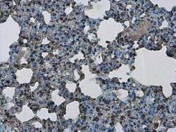
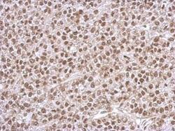
- Immunohistochemistry (Paraffin) analysis of PHF10 was performed in paraffin-embedded rat lung tissue using PHF10 Polyclonal Antibody (Product # PA5-30678) at a dilution of 1:500.
- Submitted by
- Invitrogen Antibodies (provider)
- Main image
- Experimental details
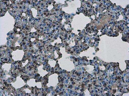
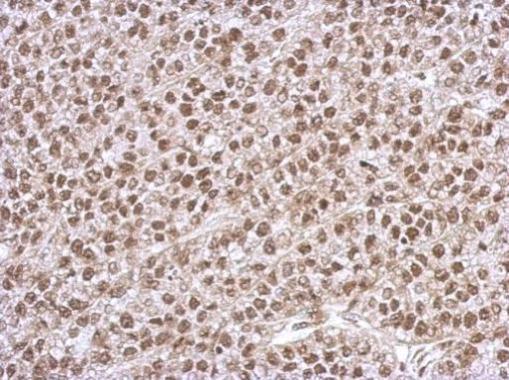
- Immunohistochemical analysis of paraffin-embedded Hela xenograft, using PHF10 (Product # PA5-30678) antibody at 1:500 dilution. Antigen Retrieval: EDTA based buffer, pH 8.0, 15 min.
Supportive validation
- Submitted by
- Invitrogen Antibodies (provider)
- Main image

- Experimental details
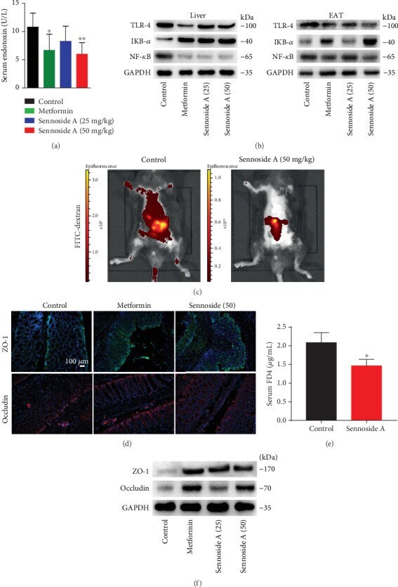
- Figure 5 Sennoside A improved intestinal integrity and metabolic endotoxemia in db / db mice. Serum endotoxin (a) and intestinal permeability (c and e) were measured as described in Materials and Methods. Effects of Sennoside A treatment on TLR4, IkB- alpha , and NF- kappa B in the liver and EAT (b). Sennoside A prevented expressions and corrected distribution of colonic tight junction proteins, occludin, and ZO-1 (scale bar: 100 mu m) (d). Western blot results were consistent with immunofluorescence results, showing a dose-dependent inhibition of expressions of the tight junction proteins in colon tissues (f).
- Submitted by
- Invitrogen Antibodies (provider)
- Main image

- Experimental details
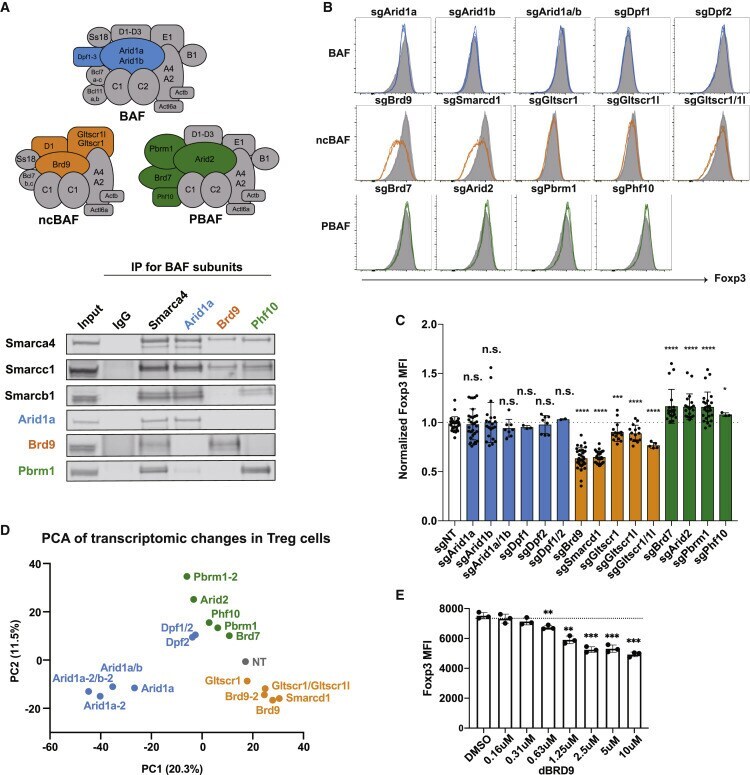
- Figure 3 The Three SWI/SNF Complex Assemblies Have Distinct Regulatory Roles for Foxp3 Expression in Treg Cells (A) A diagram showing three different variants of SWI/SNF complexes: BAF, ncBAF, and PBAF. BAF-specific subunits (Arid1a and Dpf1-Dpf3) are colored blue, ncBAF-specific subunits (Brd9, Smarcd1, Gltscr1l, and Gltscr1) are colored orange, and PBAF-specific subunits (Pbrm1, Arid2, Brd7, and Phf10) are colored green. Shared components among complexes are colored gray. Also shown is an immunoprecipitation assay of Arid1a, Brd9, Phf10, and Smarca4 in Treg cells. The co-precipitated proteins were probed for shared subunits (Smarca4, Smarcc1, and Smarcb1), BAF-specific Arid1a, ncBAF-specific Brd9, and PBAF-specific Pbrm1. (B) FACS histogram of Foxp3 expression in Treg cells after sgRNA targeting of the indicated SWI/SNF subunits. (C) MFI of Foxp3 after sgRNA targeting of the indicated SWI/SNF subunits. Data represent mean and standard deviation of biological replicates (n = 3-21). (D) Principal-component analysis of RNA-seq data collected from Treg cells transduced with guides against the indicated SWI/SNF subunits. In cases where two independent guides were used to target a gene, the second guide for targeting the gene is indicated as ""-2."" (E) MFI of Foxp3 expression in Treg cells after treatment with DMSO or 0.16-10 muM dBRD9 for 4 days. Data represent mean +- SD. Statistical analyses were performed using unpaired two-tailed Student's t test (non-significant [ns], p >=