Explore
 Validate
 Learn
Learn Western blot
Antibody data
- Antibody Data
- Antigen structure
- References [0]
- Comments [0]
- Validations
- Western blot [1]
- Immunocytochemistry [1]
- Immunohistochemistry [1]
- Flow cytometry [1]
Submit
Validation data
Reference
Comment
Report error
- Product number
- GTX80981 - Provider product page

- Provider
- GeneTex
- Proper citation
- GeneTex Cat#GTX80981, RRID:AB_11178901
- Product name
- PSMB1 antibody, C-term
- Antibody type
- Polyclonal
- Reactivity
- Human, Mouse
- Host
- Rabbit
No comments: Submit comment
Supportive validation
- Submitted by
- GeneTex (provider)
- Main image
- Experimental details
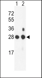
- Western blot analysis of PSMB1 Antibody (C-term) (GTX80981) in mouse NIH-3T3 cell line(lane 1) and mouse bladder tissue(lane 2) lysates (35ug/lane). PSMB1 (arrow) was detected using the purified Pab.
- Validation comment
- WB

Supportive validation
- Submitted by
- GeneTex (provider)
- Main image
- Experimental details
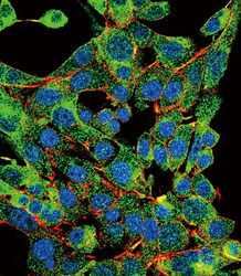
- Confocal immunofluorescent analysis of PSMB1 Antibody (C-term)(GTX80981) with HepG2 cell followed by Alexa Fluor 488-conjμgated goat anti-rabbit lgG (green). Actin filaments have been labeled with Alexa Fluor 555 phalloidin (red). DAPI was used to stain the cell nuclear (blue).

Supportive validation
- Submitted by
- GeneTex (provider)
- Main image
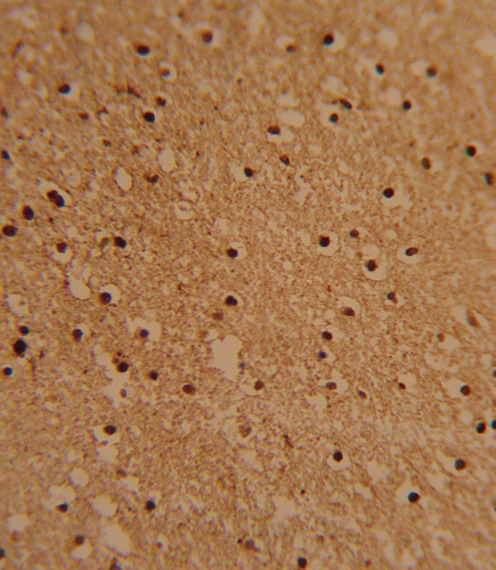
- Experimental details
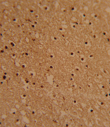
- Formalin-fixed and paraffin-embedded human brain tissue reacted with PSMB1 Antibody (C-term) (GTX80981), which was peroxidase-conjμgated to the secondary antibody, followed by DAB staining. This data demonstrates the use of this antibody for immunohistochemistry; clinical relevance has not been evaluated.
Supportive validation
- Submitted by
- GeneTex (provider)
- Main image
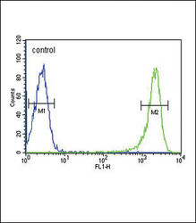
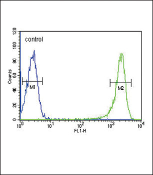
- Experimental details
- PSMB1 Antibody (C-term) (GTX80981) flow cytometric analysis of HL-60 cells (right histogram) compared to a negative control cell (left histogram).FITC-conjμgated goat-anti-rabbit secondary antibodies were used for the analysis.