Explore
 Validate
 Learn
Learn Western blot
 Immunocytochemistry
Antibody data
- Antibody Data
- Antigen structure
- References [3]
- Comments [0]
- Validations
- Immunocytochemistry [2]
- Immunohistochemistry [1]
- Other assay [3]
Submit
Validation data
Reference
Comment
Report error
- Product number
- PA3-116 - Provider product page

- Provider
- Invitrogen Antibodies
- Product name
- CCKAR Polyclonal Antibody
- Antibody type
- Polyclonal
- Antigen
- Synthetic peptide
- Description
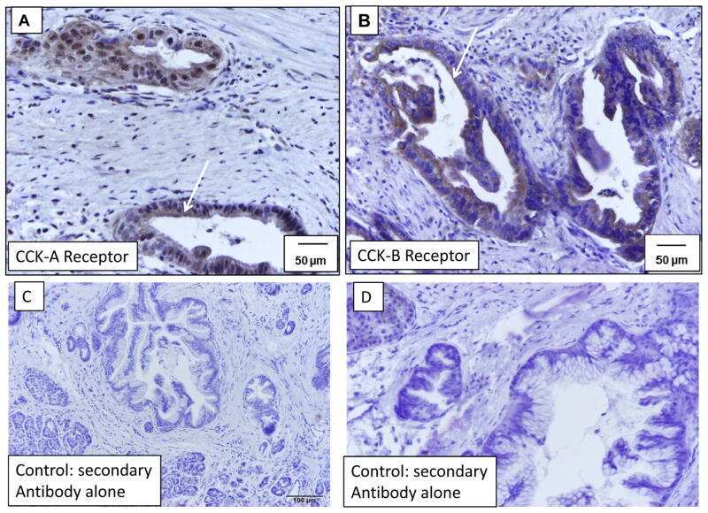
- PA3-116 detects CCK1-Receptor in human samples. PA3-116 has been successfully used in Western blot, immunocytochemistry and immunohistochemistry procedures. By Western blot, this antibody detects an ~80-90 kDa protein representing CCK1-Receptor in human samples. In immunohistochemistry, PAC1 immunoreactivity is predominantly localized at the plasma membrane and in cells of the gastric mucosa, myenteric ganglion cells, myenteric nerve fibers in the gasrtointestinal tract. PA3-116 immunogen is a synthetic peptide, KLH conjugated, corresponding to the residues T(409) G A S L S R F S Y S H M S A S V P P Q (428) of human VPAC2 receptor.
- Reactivity
- Human
- Host
- Rabbit
- Isotype
- IgG
- Vial size
- 100 μL
- Concentration
- Conc. Not Determined
- Storage
- -20°C, Avoid Freeze/Thaw Cycles
Submitted references Cholecystokinin receptor antagonist halts progression of pancreatic cancer precursor lesions and fibrosis in mice.
Cholecystokinin mediates progression and metastasis of pancreatic cancer associated with dietary fat.
Immunohistochemical localization of CCK1 cholecystokinin receptors in normal and neoplastic human tissues.
Smith JP, Cooper TK, McGovern CO, Gilius EL, Zhong Q, Liao J, Molinolo AA, Gutkind JS, Matters GL
Pancreas 2014 Oct;43(7):1050-9
Pancreas 2014 Oct;43(7):1050-9
Cholecystokinin mediates progression and metastasis of pancreatic cancer associated with dietary fat.
Matters GL, Cooper TK, McGovern CO, Gilius EL, Liao J, Barth BM, Kester M, Smith JP
Digestive diseases and sciences 2014 Jun;59(6):1180-91
Digestive diseases and sciences 2014 Jun;59(6):1180-91
Immunohistochemical localization of CCK1 cholecystokinin receptors in normal and neoplastic human tissues.
Schulz S, Röcken C, Mawrin C, Schulz S
The Journal of clinical endocrinology and metabolism 2005 Nov;90(11):6149-55
The Journal of clinical endocrinology and metabolism 2005 Nov;90(11):6149-55
No comments: Submit comment
Supportive validation
- Submitted by
- Invitrogen Antibodies (provider)
- Main image

- Experimental details
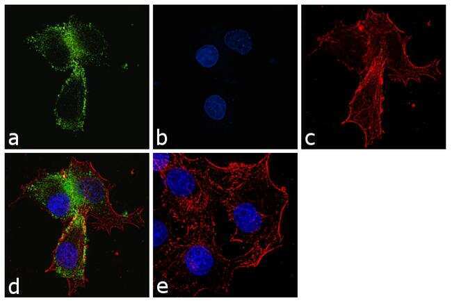
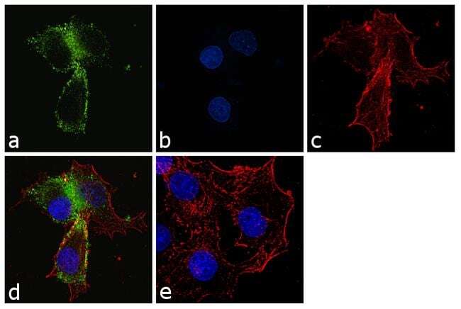
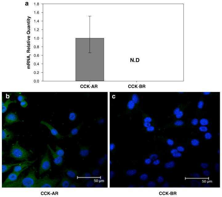
- Immunofluorescence analysis of CCK-A Receptor was performed using 70% confluent log phase PANC-1 cells. The cells were fixed with 4% paraformaldehyde for 10 minutes, permeabilized with 0.1% Triton™ X-100 for 10 minutes, and blocked with 1% BSA for 1 hour at room temperature. The cells were labeled with CCKAR Rabbit Polyclonal Antibody (Product # PA3-116) at 1:250 dilution in 0.1% BSA and incubated for 3 hours at room temperature and then labeled with Goat anti-Rabbit IgG (H+L) Superclonal™ Secondary Antibody, Alexa Fluor® 488 conjugate (Product # A27034) at a dilution of 1:2000 for 45 minutes at room temperature (Panel a: green). Nuclei (Panel b: blue) were stained with SlowFade® Gold Antifade Mountant with DAPI (Product # S36938). F-actin (Panel c: red) was stained with Rhodamine Phalloidin (Product # R415, 1:300). Panel d represents the merged image showing membranous localization. Panel e shows the no primary antibody control. The images were captured at 60X magnification.
- Submitted by
- Invitrogen Antibodies (provider)
- Main image
- Experimental details
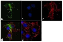
- Immunofluorescence analysis of CCK-A Receptor was performed using 70% confluent log phase PANC-1 cells. The cells were fixed with 4% paraformaldehyde for 10 minutes, permeabilized with 0.1% Triton™ X-100 for 10 minutes, and blocked with 1% BSA for 1 hour at room temperature. The cells were labeled with CCKAR Rabbit Polyclonal Antibody (Product # PA3-116) at 1:250 dilution in 0.1% BSA and incubated for 3 hours at room temperature and then labeled with Goat anti-Rabbit IgG (Heavy Chain) Superclonal™ Secondary Antibody, Alexa Fluor® 488 conjugate (Product # A27034) at a dilution of 1:2000 for 45 minutes at room temperature (Panel a: green). Nuclei (Panel b: blue) were stained with SlowFade® Gold Antifade Mountant with DAPI (Product # S36938). F-actin (Panel c: red) was stained with Rhodamine Phalloidin (Product # R415, 1:300). Panel d represents the merged image showing membranous localization. Panel e shows the no primary antibody control. The images were captured at 60X magnification.
Supportive validation
- Submitted by
- Invitrogen Antibodies (provider)
- Main image
- Experimental details
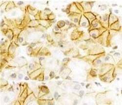
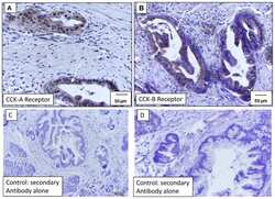
- Immunohistochemical staining of CCK1-Receptor in human gastric mucosa samples using Product # PA3-116.

Supportive validation
- Submitted by
- Invitrogen Antibodies (provider)
- Main image

- Experimental details
- NULL
- Submitted by
- Invitrogen Antibodies (provider)
- Main image
- Experimental details
- NULL
- Submitted by
- Invitrogen Antibodies (provider)
- Main image
- Experimental details
- NULL