 Explore
 Validate
 Learn
LearnMAB36254-100
antibody from R&D Systems
Targeting: IL33
C9orf26, DKFZp586H0523, DVS27, IL1F11, NF-HEV
 Western blot
Antibody data
- Antibody Data
- Antigen structure
- References [0]
- Comments [0]
- Validations
- Western blot [1]
- Immunohistochemistry [1]
- Blocking/Neutralizing [1]
Submit
Validation data
Reference
Comment
Report error
- Product number
- MAB36254-100 - Provider product page

- Provider
- R&D Systems
- Product name
- Human IL-33 Antibody
- Antibody type
- Monoclonal
- Description
- Protein A or G purified from cell culture supernatant. Detects human IL-33 in direct ELISAs.
- Reactivity
- Human
- Host
- Goat
- Conjugate
- Unconjugated
- Antigen sequence
O95760- Isotype
- IgG
- Antibody clone number
- 40015D
- Vial size
- 100 ug
- Storage
- Use a manual defrost freezer and avoid repeated freeze-thaw cycles. 12 months from date of receipt, -20 to -70 °C as supplied. 1 month, 2 to 8 °C under sterile conditions after reconstitution. 6 months, -20 to -70 °C under sterile conditions after reconstitution.
No comments: Submit comment
Supportive validation
- Submitted by
- R&D Systems (provider)
- Main image

- Experimental details
- Detection of Human IL-33 by Western Blot. Western blot shows lysates of HEK293 human embryonic kidney cell line either mock transfected or transfected with human IL-33. PVDF membrane was probed with 1 µg/mL of Recombinant Goat Anti-Human IL-33 Monoclonal Antibody (Catalog # MAB36254) followed by HRP-conjugated Anti-Goat IgG Secondary Antibody (Catalog # HAF017). A specific band was detected for IL-33 at approximately 30 kDa (as indicated). This experiment was conducted under reducing conditions and using Immunoblot Buffer Group 1.

Supportive validation
- Submitted by
- R&D Systems (provider)
- Main image
- Experimental details
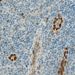
- IL-33 in Human Tonsil. IL-33 was detected in immersion fixed paraffin-embedded sections of human tonsil using Recombinant Goat Anti-Human IL-33 Monoclonal Antibody (Catalog # MAB36254) at 0.3 µg/mL overnight at 4 °C. Tissue was stained using the Anti-Goat HRP-DAB Cell & Tissue Staining Kit (brown; Catalog # CTS008) and counterstained with hematoxylin (blue). Specific staining was localized to nuclei. View our protocol for Chromogenic IHC Staining of Paraffin-embedded Tissue Sections.

Supportive validation
- Submitted by
- R&D Systems (provider)
- Main image

- Experimental details
- Cell Proliferation Induced by IL-33 and Neutralization by Human IL-33 Antibody. In the presence of sub-optimal amounts of Hamster Anti-Mouse CD3 epsilon Monoclonal Antibody (Catalog # MAB484), Recombinant Human IL-33 (Catalog # 3625-IL) stimulates proliferation in the D10.G4.1 mouse helper T cell line in a dose-dependent manner (orange line), as measured by Resazurin (Catalog # AR002). Under these conditions, proliferation elicited by Recombinant Human IL-33 (12 ng/mL) is neutralized (green line) by increasing concentrations of Goat Anti-Human IL-33 Monoclonal Antibody (Catalog # MAB36254). The ND50 is typically 0.75-3 µg/mL.
