Explore
 Validate
 Learn
LearnMAB36253-100
antibody from R&D Systems
Targeting: IL33
C9orf26, DKFZp586H0523, DVS27, IL1F11, NF-HEV
 Western blot
 ELISA
ELISAAntibody data
- Antibody Data
- Antigen structure
- References [1]
- Comments [0]
- Validations
- Western blot [1]
- Immunohistochemistry [1]
- Flow cytometry [1]
Submit
Validation data
Reference
Comment
Report error
- Product number
- MAB36253-100 - Provider product page

- Provider
- R&D Systems
- Product name
- Human IL-33 Antibody
- Antibody type
- Monoclonal
- Description
- Protein A or G purified from cell culture supernatant. Detects human IL-33 in direct ELISAs and Western blots.
- Reactivity
- Human
- Host
- Goat
- Conjugate
- Unconjugated
- Antigen sequence
O95760- Isotype
- IgG
- Antibody clone number
- 40015C
- Vial size
- 100 ug
- Storage
- Use a manual defrost freezer and avoid repeated freeze-thaw cycles. 12 months from date of receipt, -20 to -70 °C as supplied. 1 month, 2 to 8 °C under sterile conditions after reconstitution. 6 months, -20 to -70 °C under sterile conditions after reconstitution.
Submitted references Serial Monitoring of Immune Markers Being Represented Regulatory T Cell/T Helper 17 Cell Ratio: Indicating Tolerance for Tapering Immunosuppression after Liver Transplantation.
Jhun J, Lee SH, Lee SK, Kim HY, Jung ES, Kim DG, Choi J, Bae SH, Yoon SK, Chung BH, Yang CW, Cho ML, Choi JY
Frontiers in immunology 2018;9:352
Frontiers in immunology 2018;9:352
No comments: Submit comment
Supportive validation
- Submitted by
- R&D Systems (provider)
- Main image
- Experimental details
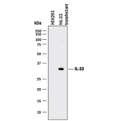
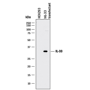
- Detection of Human IL-33 by Western Blot. Western blot shows lysates of HEK293 human embryonic kidney cell line either mock transfected or transfected with human IL-33. PVDF membrane was probed with 1 µg/mL of Goat Anti-Human IL-33 Monoclonal Antibody (Catalog # MAB36253) followed by HRP-conjugated Anti-Goat IgG Secondary Antibody (Catalog # HAF017). A specific band was detected for IL-33 at approximately 30 kDa (as indicated). This experiment was conducted under reducing conditions and using Immunoblot Buffer Group 1.
Supportive validation
- Submitted by
- R&D Systems (provider)
- Main image
- Experimental details
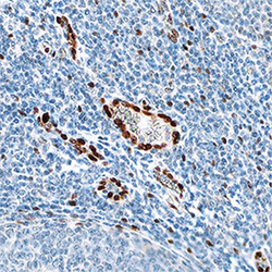
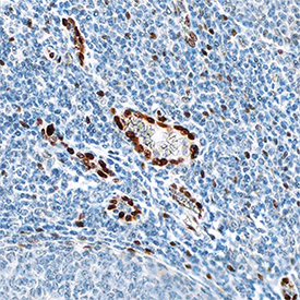
- IL-33 in Human Tonsil. IL-33 was detected in immersion fixed paraffin-embedded sections of human tonsil using Goat Anti-Human IL-33 Monoclonal Antibody (Catalog # MAB36253) at 0.1 µg/mL overnight at 4 °C. Tissue was stained using the Anti-Goat HRP-DAB Cell & Tissue Staining Kit (brown; Catalog # CTS008) and counterstained with hematoxylin (blue). Specific staining was localized to nuclei in epithelial cells. View our protocol for Chromogenic IHC Staining of Paraffin-embedded Tissue Sections.
Supportive validation
- Submitted by
- R&D Systems (provider)
- Main image

- Experimental details
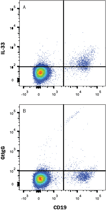
- Detection of IL-33 in Human Peripheral Blood Lymphocytes by Flow Cytometry. Human peripheral blood lymphocytes were stained with (A) Goat Anti-Human IL-33 Monoclonal Antibody (Catalog # MAB36253) or (B) Goat IgG control antibody (Catalog # AB-108-C) followed by anti-Goat IgG PE-conjugated secondary antibody (Catalog # F0107) and Mouse anti-Human CD19 APC-conjugated Monoclonal Antibody (Catalog # FAB4867A). To facilitate intracellular staining, cells were fixed with Flow Cytometry Fixation Buffer (Catalog # FC004) and permeabilized with methanol. View our protocol for Staining Intracellular Molecules.