 Explore
 Validate
 Learn
LearnPAB16924
antibody from Abnova Corporation
Targeting: IL33
C9orf26, DKFZp586H0523, DVS27, IL1F11, NF-HEV
 Western blot
Antibody data
- Antibody Data
- Antigen structure
- References [3]
- Comments [0]
- Validations
- Western blot [1]
Submit
Validation data
Reference
Comment
Report error
- Product number
- PAB16924 - Provider product page

- Provider
- Abnova Corporation
- Proper citation
- Abnova Corporation Cat#PAB16924, RRID:AB_10678330
- Product name
- IL33 polyclonal antibody
- Antibody type
- Polyclonal
- Description
- Rabbit polyclonal antibody raised against full length recombinant IL33.
- Storage
- Store at 4°C on dry atmosphere.After reconstitution with 0.1 mL of deionized water, store at -20°C or lower.Aliquot to avoid repeated freezing and thawing.
Submitted references Mature interleukin-33 is produced by calpain-mediated cleavage in vivo.
IL-33, a recently identified interleukin-1 gene family member, is expressed in human adipocytes.
Suppression of interleukin-33 bioactivity through proteolysis by apoptotic caspases.
Hayakawa M, Hayakawa H, Matsuyama Y, Tamemoto H, Okazaki H, Tominaga S
Biochemical and biophysical research communications 2009 Sep 11;387(1):218-22
Biochemical and biophysical research communications 2009 Sep 11;387(1):218-22
IL-33, a recently identified interleukin-1 gene family member, is expressed in human adipocytes.
Wood IS, Wang B, Trayhurn P
Biochemical and biophysical research communications 2009 Jun 19;384(1):105-9
Biochemical and biophysical research communications 2009 Jun 19;384(1):105-9
Suppression of interleukin-33 bioactivity through proteolysis by apoptotic caspases.
Lüthi AU, Cullen SP, McNeela EA, Duriez PJ, Afonina IS, Sheridan C, Brumatti G, Taylor RC, Kersse K, Vandenabeele P, Lavelle EC, Martin SJ
Immunity 2009 Jul 17;31(1):84-98
Immunity 2009 Jul 17;31(1):84-98
No comments: Submit comment
Supportive validation
- Submitted by
- Abnova Corporation (provider)
- Main image
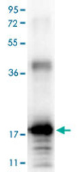
- Experimental details
- Western blot using IL33 polyclonal antibody (Cat # PAB16924) shows detection of a band ~18 KDa in size corresponding to recombinant human IL33. After transfer, the membrane was blocked overnight with 3% BSA in TBS followed by reaction with primary antibody at a 1 : 1,000 dilution. Detection occurred using peroxidase conjugated anti-Rabbit IgG secondary antibody diluted 1 : 40,000 in blocking buffer for 30 min at RT followed by reaction with FemtoMax™ chemiluminescent substrate. Image was captured using VersaDoc™ MP 4000 imaging system (Bio-Rad).
