 Explore
 Validate
 Learn
Learn Western blot
Antibody data
- Antibody Data
- Antigen structure
- References [4]
- Comments [0]
- Validations
- Western blot [4]
- Immunocytochemistry [1]
- Other assay [3]
Submit
Validation data
Reference
Comment
Report error
- Product number
- PA5-17256 - Provider product page

- Provider
- Invitrogen Antibodies
- Product name
- MAVS Polyclonal Antibody
- Antibody type
- Polyclonal
- Antigen
- Synthetic peptide
- Description
- It is not recommended to aliquot this antibody.
- Reactivity
- Human
- Host
- Rabbit
- Isotype
- IgG
- Vial size
- 100 µL
- Concentration
- 71 µg/mL
- Storage
- -20°C
Submitted references CD169-mediated restrictive SARS-CoV-2 infection of macrophages induces pro-inflammatory responses.
The microRNAs miR-302b and miR-372 regulate mitochondrial metabolism via the SLC25A12 transporter, which controls MAVS-mediated antiviral innate immunity.
HIV-1 intron-containing RNA expression induces innate immune activation and T cell dysfunction.
Viral suppressors of the RIG-I-mediated interferon response are pre-packaged in influenza virions.
Jalloh S, Olejnik J, Berrigan J, Nisa A, Suder EL, Akiyama H, Lei M, Tyagi S, Bushkin Y, Mühlberger E, Gummuluru S
bioRxiv : the preprint server for biology 2022 Mar 30;
bioRxiv : the preprint server for biology 2022 Mar 30;
The microRNAs miR-302b and miR-372 regulate mitochondrial metabolism via the SLC25A12 transporter, which controls MAVS-mediated antiviral innate immunity.
Yasukawa K, Kinoshita D, Yaku K, Nakagawa T, Koshiba T
The Journal of biological chemistry 2020 Jan 10;295(2):444-457
The Journal of biological chemistry 2020 Jan 10;295(2):444-457
HIV-1 intron-containing RNA expression induces innate immune activation and T cell dysfunction.
Akiyama H, Miller CM, Ettinger CR, Belkina AC, Snyder-Cappione JE, Gummuluru S
Nature communications 2018 Aug 27;9(1):3450
Nature communications 2018 Aug 27;9(1):3450
Viral suppressors of the RIG-I-mediated interferon response are pre-packaged in influenza virions.
Liedmann S, Hrincius ER, Guy C, Anhlan D, Dierkes R, Carter R, Wu G, Staeheli P, Green DR, Wolff T, McCullers JA, Ludwig S, Ehrhardt C
Nature communications 2014 Dec 9;5:5645
Nature communications 2014 Dec 9;5:5645
No comments: Submit comment
Supportive validation
- Submitted by
- Invitrogen Antibodies (provider)
- Main image

- Experimental details
- Knockout of MAVS was achieved by CRISPR-Cas9 genome editing using LentiArray™ Lentiviral sgRNA (Product # A32042, Assay ID CRISPR988932_LV) and LentiArray Cas9 Lentivirus (Product # A32064). Western blot analysis of MAVS was performed by loading 60 µg of 293-FT wild type (Lane 1), 293-FT Cas9 (Lane 2) and 293-FT MAVS KO (Lane 3) whole cell extracts. The samples were electrophoresed using NuPAGE™ Novex™ 4-12% Bis-Tris Protein Gel (Product # NP0322BOX). Resolved proteins were then transferred onto a nitrocellulose membrane (Product # IB23001) by iBlot® 2 Dry Blotting System (Product # IB21001). The blot was probed with MAVS Polyclonal Antibody (Product # PA5-17256, 1:1000 dilution) and Goat anti-Rabbit IgG (H+L) Superclonal™ Recombinant Secondary Antibody, HRP (Product # A27036, 1:12,000 dilution) with the iBright™ FL1500 (Product # A44115). Chemiluminescent detection was performed using SuperSignal™ West Dura Extended Duration Substrate (Product # 34076) with the iBright™ FL1500 (Product # A44115). Loss of signal upon CRISPR mediated knockout (KO) using the LentiArray™ CRISPR product line confirms that antibody is specific to MAVS. Uncharacterized band was observed in all the samples at ~50 kDa.

- Submitted by
- Invitrogen Antibodies (provider)
- Main image

- Experimental details
- Western blot analysis of MAVS in extracts from MCF-7 and HT29 cell lines using MAVS polyclonal antibody (Product # PA5-17256).

- Submitted by
- Invitrogen Antibodies (provider)
- Main image

- Experimental details
- Western blot analysis of MAVS in extracts from HeLa cells, mock-transfected or transfected with human MAVS, using MAVS polyclonal antibody (Product # PA5-17256).

- Submitted by
- Invitrogen Antibodies (provider)
- Main image
- Experimental details
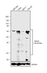
- Western blot was performed using Anti-MAVS Polyclonal Antibody(Product # PA5-17256) and a 60-30kDa band corresponding to MAVS was observed across cell lines tested. Membrane enriched extracts (30 µg lysate) of HEK-293 (Lane 1), A549 (Lane 2), MCF-7 (Lane 3) and Hep G2 (Lane 4) were electrophoresed using NuPAGE™ 4-12% Bis-Tris Protein Gel (Product # NP0322BOX). Resolved proteins were then transferred onto a Nitrocellulose membrane (Product # IB23001) by iBlot® 2 Dry Blotting System (Product # IB21001). The blot was probed with the primary antibody (1:1000) and detected by chemiluminescence with Goat anti-Rabbit IgG (H+L) Superclonal™ Recombinant Secondary Antibody, HRP (Product # A27036,1:4000) using the iBright FL 1000 (Product # A32752). Chemiluminescent detection was performed using SuperSignal™ West Dura Extended Duration Substrate (Product # 34076).

Supportive validation
- Submitted by
- Invitrogen Antibodies (provider)
- Main image

- Experimental details
- Immunofluorescent analysis of MAVS in MCF-7 cells using a MAVS polyclonal antibody (Product # PA5-17256) (green) showing colocalization with mitochondria that have been labeled with a mitochondrial fluorescent red dye. DNA is labeled using a fluorescent blue dye.

Supportive validation
- Submitted by
- Invitrogen Antibodies (provider)
- Main image

- Experimental details
- Fig. 5 MDM immune activation is initiated via a MAVS-dependent pathway. a MDMs were transduced with lentivectors expressing shRNA against scrambled sequence (Sc), MAVS or STING, and the expression of host proteins targeted by shRNAs was analyzed by immune blotting. b - e shRNA-transduced MDMs were infected with HIV-1 (Lai[delta]envGFP/G) and viral expression (GFP) ( b , c ) and CD169 expression ( d , e ) were analyzed 6 days post infection. f MDMs were transduced with lentivectors expressing shRNA against scrambled sequence (Sc), RIG-I or MDA5, and the expression host proteins targeted by shRNAs analyzed by immune blotting. g - j shRNA-transduced MDMs were infected with Lai[delta]envGFP/G and viral expression (GFP) ( g , h ) and CD169 expression ( i , j ) were analyzed 6 days post infection. k - m Effect of inhibitors on HIV-1 infection ( k ), CD169 expression ( l ), and IP-10 production ( m ) in MDMs. MDMs were infected with HIV-1 (MOI of 2) and cultured in the presence of indicated inhibitors for 6 days. mock1/DMSO1: 0.1% DMSO, mock2/DMSO2: 0.01% DMSO. The means +- SEM are shown and each symbol represents an independent experiment. Two-tailed p -values: one-way ANOVA followed by the Tukey-Kramer post-test ( d , e , i , j , l , and m ) or paired t -test ( b , c , g , and h ). * p < 0.05, ** p < 0.01, *** p < 0.001, ns: not significant

- Submitted by
- Invitrogen Antibodies (provider)
- Main image

- Experimental details
- Figure 1 High-resolution visualization of internalized IAV vRNPs revealed helical-like structure in close proximity to RIG-I and MAVS at the mitochondrial membrane. ( a - e ) A549 cells were infected with H1N1-WT (multiplicity of infection (MOI)=50) and prepared for immunofluorescence analysis 3 h post infection (h.p.i.). ( a - c ) Three-dimensional stochastic optical reconstruction microscopy (STORM) revealed internalized vRNPs (green: NP-AF568) as helical-like structure ( Supplementary Movie 1 ) in close association with the outer mitochondrial membrane (red: TOM20-AF647). ( a ) Wide shot, scale bar represents 5 mum, ( b ) zoom in, scale bar represents 200 nm and ( c ) zoom in 3D render. ( d , e ) vRNPs (green: NP-atto488) are in close proximity to ( d ) RIG-I (cyan: RIG-I-AF568) and ( e ) MAVS (cyan: MAVS-AF568) at the outer mitochondrial membrane (red: TOM20-biotin-streptavidin-AF647). Shown are representative images from three independent experiments each with at least 40 analysed cells. ( f ) Model of a mitochondria-associated vRNP. Coloured shapes indicate structure visualized in STORM analysis.

- Submitted by
- Invitrogen Antibodies (provider)
- Main image

- Experimental details
- Figure 7. MAVS is essential for SARS-CoV-2 RNA-induced inflammatory responses in macrophages. ( A ) Parental (THP1) monocytes and those expressing CD169, ACE2, or CD169/ACE2 were transduced with lentivectors expressing shRNA against scrambled sequence (-), or MAVS sequence (+), and knockdown of MAVS in each cell line analyzed by immune blotting ( A , left panel), and quantified ( A , right panel). ( B-C ) THP1 monocytes with stable MAVS knockdown were PMA-differentiated and infected with SARS-CoV-2 (MOI=1, 24 hpi), and total RNA analyzed by RT-qPCR for ( B ) total viral transcripts (nucleocapsid), and ( C ) IL6, TNFalpha, IL-1beta and IFNlambda1 mRNA expression, normalized to mock-infected controls for each group. The means +- SEM from 3 independent experiments are shown, and significant differences were determined by one-way ANOVA followed by the Tukey-Kramer post-test within groups ( B , and C ), or Dunnett's post-test between groups ( C ). P -values: *
