Explore
 Validate
 Learn
LearnPA5-40774
antibody from Invitrogen Antibodies
Targeting: TIPARP
ARTD14, DDF1, DKFZP434J214, DKFZp686N0351, PARP-1, PARP-7, PARP7, pART14, RM1
 Western blot
Antibody data
- Antibody Data
- Antigen structure
- References [1]
- Comments [0]
- Validations
- Western blot [1]
- Other assay [5]
Submit
Validation data
Reference
Comment
Report error
- Product number
- PA5-40774 - Provider product page

- Provider
- Invitrogen Antibodies
- Product name
- TIPARP Polyclonal Antibody
- Antibody type
- Polyclonal
- Antigen
- Synthetic peptide
- Description
- Peptide sequence: LKTCFKKKDQ KRLGTGTLRS LRPILNTLLE SGSLDGVFRS RNQSTDENSL Sequence homology: Cow: 100%; Dog: 100%; Horse: 100%; Human: 100%; Mouse: 83%; Pig: 100%; Rabbit: 92%; Rat: 83%
- Reactivity
- Human
- Host
- Rabbit
- Isotype
- IgG
- Vial size
- 100 μL
- Concentration
- 0.5 mg/mL
- Storage
- -20°C, Avoid Freeze/Thaw Cycles
Submitted references Identification of PARP-7 substrates reveals a role for MARylation in microtubule control in ovarian cancer cells.
Palavalli Parsons LH, Challa S, Gibson BA, Nandu T, Stokes MS, Huang D, Lea JS, Kraus WL
eLife 2021 Jan 21;10
eLife 2021 Jan 21;10
No comments: Submit comment
Supportive validation
- Submitted by
- Invitrogen Antibodies (provider)
- Main image
- Experimental details
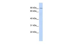
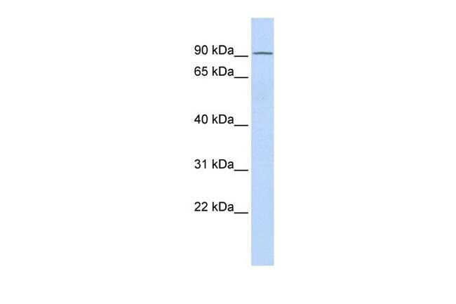
- Western blot analysis of human heart cells using an anti-TIPARP polyclonal antibody (Product # PA5-40774).
Supportive validation
- Submitted by
- Invitrogen Antibodies (provider)
- Main image
- Experimental details
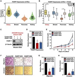
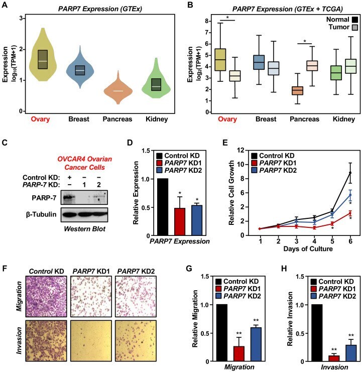
- Figure 1. PARP-7 expression in cancers and role in ovarian cancer cell phenotypes. ( A ) Violin plots showing normalized expression of PARP7 mRNA from GTEx data presented as log 10 (TPM+1) (Transcripts Per Million), calculated from a gene model with all isoforms collapsed to a single gene. The data were obtained from the GTEx portal and expressed as normalized TPM scores as described (). Expression profiles of PARP7 mRNA in the following cancers: breast (n = 459, median TPM = 21.1), ovary (n = 180, median TPM = 37.6), pancreas (n = 328, median TPM = 4.1), and kidney cortex (n = 85, median TPM = 5.6). ( B ) Box plots showing normalized expression of PARP7 mRNA across four tissues (ovary, breast, pancreas, and kidney) and their corresponding cancer types (matched TCGA normal and GTEx) analyzed using GEPIA and presented as log 2 (TPM+1). Ovary and pancreas show significantly different expression levels for PARP7 between normal and tumor tissues. Asterisks indicate significant differences between normal and tumor samples (Student's t-test, *p
- Submitted by
- Invitrogen Antibodies (provider)
- Main image
- Experimental details
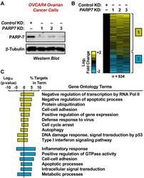
- Figure 2. RNA-seq analysis of gene expression in ovarian cancer cells following PARP-7 depletion. ( A ) Western blots showing PARP-7 protein levels after siRNA-mediated knockdown (KD) of PARP7 in OVCAR4 cells. Three different siRNAs were used. beta-tubulin was used as loading control. ( B ) Heat maps showing the results of RNA-seq assays from OVCAR4 cells upon siRNA-mediated knockdown (KD) of PARP7. Three different siRNAs were used. Results represent fold changes in FPKM values for genes significantly regulated versus the control KD, expressed as log 2 fold change. A fold change >=1.5 was classified as upregulated, while a fold change

- Submitted by
- Invitrogen Antibodies (provider)
- Main image
- Experimental details
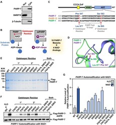
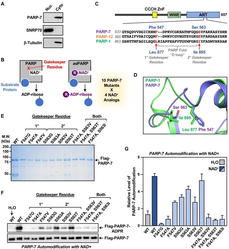
- Figure 3. Generation of NAD + analog-sensitive PARP-7 (asPARP-7) mutants. ( A ) PARP-7 localizes to the cytosolic compartment in OVCAR4 cells. Western blots of nuclear and cytosolic fractions of OVCAR4 ovarian cancer cells. SNRP70 and beta-tubulin were used as loading controls for the nuclear and cytoplasmic fractions, respectively. ( B ) Schematic diagram illustrating the NAD + analog-sensitive PARP approach. R, unnatural chemical moieties added to NAD + . In this study, 10 PARP-7 mutants targeting two potential gatekeeper residues (shown in red) were screened with 14 different NAD + analogs. ( C ) ( Top ) Schematic diagram of PARP-7 showing the functional domains, including the Cys 3 His 1 zinc finger (CCCH ZnF), the WWE PAR-binding domain (WWE), and the ADP-ribosyl transferase domain (ART). ( Bottom ) Multiple sequence alignment of PARP-7 (Q7Z3E1), PARP-1 (P09874) and PARP-12 (Q9H0J9). The two potential gatekeeper residues in PARP-7 selected for mutation are highly conserved among PARP-7, PARP-1, and PARP-12. Residues F547 and S563 in PARP-7, as well as the corresponding residues in PARP-1 (residues L877 and I895) and PARP-12 (residues F579 and S595), are indicated in red. ( D ) Close-up view of residues F547 and S563 in PARP-7. The structure was generated by comparison of available structures of PARP-1 (PDB: 3PAX) and PARP-12 (PDB: 2PQF). The molecular graphic was generated with Pymol. ( E ) Recombinant PARP-7 proteins used for asPARP-7 activity screening. SDS-PAGE analys
- Submitted by
- Invitrogen Antibodies (provider)
- Main image
- Experimental details
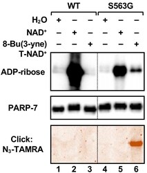
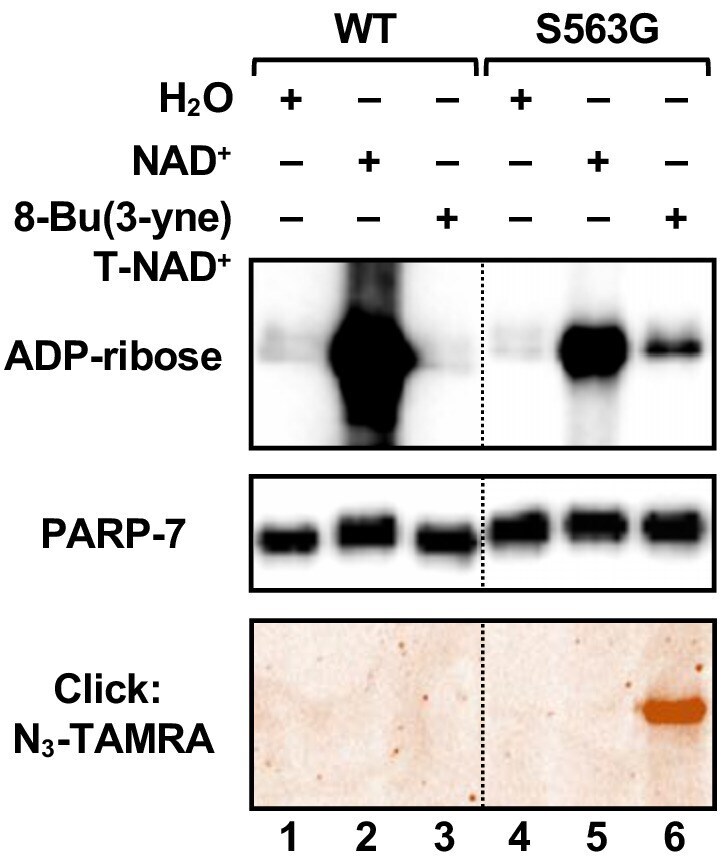
- Figure 5. Activity of the S563G analog-sensitive PARP-7 mutant with 8-Bu(3-yne)T-NAD + . Automodification reactions with wild-type and S563G mutant PARP-7 performed with NAD + or 8-Bu(3-yne)T-NAD + . The autoMARylation signals were detected by ( top ) western blotting using a MAR binding reagent or ( bottom ) click chemistry-based in-gel fluorescence. PARP-7, detected by western blotting ( middle ), was used as loading control for the reactions. TAMRA, tetramethylrhodamine.
- Submitted by
- Invitrogen Antibodies (provider)
- Main image
- Experimental details
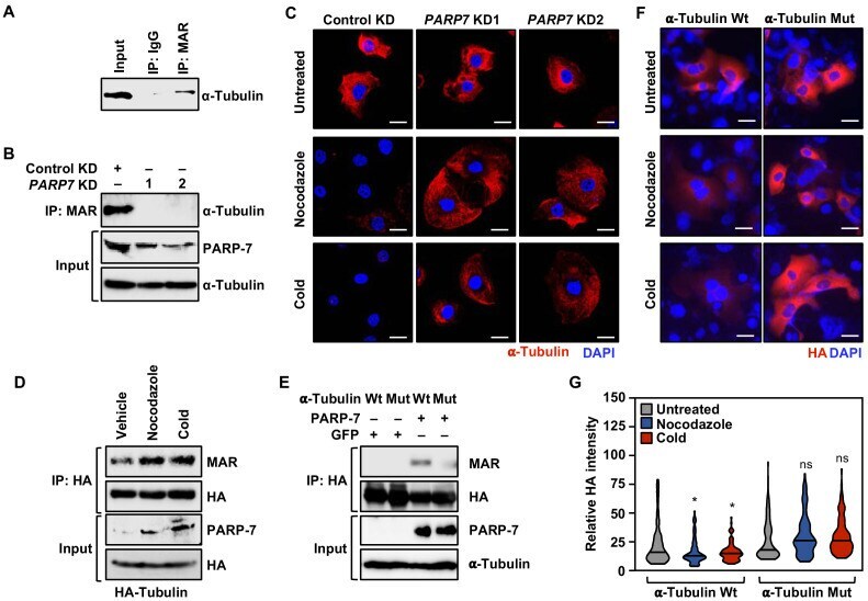
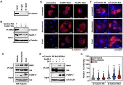
- Figure 7. PARP-7 links MARylation of alpha-tubulin to the regulation of microtubule stability in ovarian cancer cells. ( A ) MARylation of alpha-tubulin in OVCAR4 cells. MARylated proteins were immunoprecipitated from OVCAR4 cells using a MAR detection reagent and subjected to western blotting for alpha-tubulin. The experiment was performed three times (n = 3) to ensure reproducibility. ( B ) Knockdown of PARP-7 reduces the MARylation of alpha-tubulin. MARylated proteins were immunoprecipitated from OVCAR4 cells after siRNA-mediated knockdown (KD) of PARP7 and the subjected to western blotting for alpha-tubulin. The experiment was performed three times (n = 3) to ensure reproducibility. ( C ) Knockdown of PARP-7 promotes microtubule stability in OVCAR4 cells. Immunofluorescent staining of alpha-tubulin in OVCAR4 cells after siRNA-mediated knockdown (KD) of PARP7 and treatment with cold or nocodazole. Scale bars = 25 um. The experiment was performed three times (n = 3) to ensure reproducibility. ( D ) Cold or nocodazole treatment increases MARylation of alpha-tubulin. HA-tagged alpha-tubulin was immunoprecipitated from OVCAR4 cells subjected to cold or nocodazole treatment using an HA antibody. The IP material was subjected to western blotting for MAR and HA, and the input material was subjected to western blotting for PARP-7 and HA. ( E ) Ectopic expression of PARP-7 enhances the MARylation of alpha-tubulin. Wild-type (Wt) or MAR-deficient mutant alpha-tubulin was immunopreci