 Explore
 Validate
 Learn
Learn Western blot
Antibody data
- Antibody Data
- Antigen structure
- References [2]
- Comments [0]
- Validations
- Western blot [4]
- Immunocytochemistry [2]
- Immunohistochemistry [2]
Submit
Validation data
Reference
Comment
Report error
- Product number
- GTX101821 - Provider product page

- Provider
- GeneTex
- Proper citation
- GeneTex Cat#GTX101821, RRID:AB_1951757
- Product name
- RPL5 antibody
- Antibody type
- Polyclonal
- Reactivity
- Human, Mouse, Rat
- Host
- Rabbit
Submitted references Viral unmasking of cellular 5S rRNA pseudogene transcripts induces RIG-I-mediated immunity.
Splicing-factor oncoprotein SRSF1 stabilizes p53 via RPL5 and induces cellular senescence.
Chiang JJ, Sparrer KMJ, van Gent M, Lässig C, Huang T, Osterrieder N, Hopfner KP, Gack MU
Nature immunology 2018 Jan;19(1):53-62
Nature immunology 2018 Jan;19(1):53-62
Splicing-factor oncoprotein SRSF1 stabilizes p53 via RPL5 and induces cellular senescence.
Fregoso OI, Das S, Akerman M, Krainer AR
Molecular cell 2013 Apr 11;50(1):56-66
Molecular cell 2013 Apr 11;50(1):56-66
No comments: Submit comment
Supportive validation
- Submitted by
- GeneTex (provider)
- Main image
- Experimental details
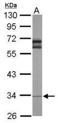
- Sample (50 ?g of whole cell lysate) A: Mouse brain 10% SDS PAGE GTX101821 diluted at 1:1000 The HRP-conjugated anti-rabbit IgG antibody (GTX213110-01) was used to detect the primary antibody.

- Submitted by
- GeneTex (provider)
- Main image

- Experimental details
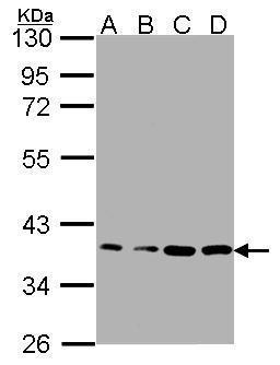
- Sample(30 ?g whole cell lysate)A: A431 (GTX27909)B: H1299 C: HeLaS3 D: MOLT4 (GTX27912)12% SDS PAGEGTX101821 diluted at 1:1000The HRP-conjugated anti-rabbit IgG antibody (GTX213110-01) was used to detect the primary antibody.
- Submitted by
- GeneTex (provider)
- Main image

- Experimental details
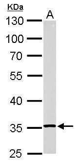
- RPL5 antibody detects RPL5 protein by western blot analysis.A. 30 ?g Rat2 whole cell lysate/extract10% SDS-PAGERPL5 antibody (GTX101821) dilution: 1:1000 The HRP-conjugated anti-rabbit IgG antibody (GTX213110-01) was used to detect the primary antibody.
- Submitted by
- GeneTex (provider)
- Main image
- Experimental details
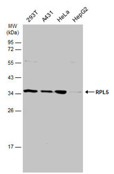
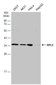
- Various whole cell extracts (30 ?g) were separated by 12% SDS-PAGE, and the membrane was blotted with RPL5 antibody (GTX101821) diluted at 1:1000. The HRP-conjugated anti-rabbit IgG antibody (GTX213110-01) was used to detect the primary antibody.
Supportive validation
- Submitted by
- GeneTex (provider)
- Main image
- Experimental details
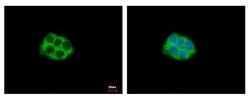
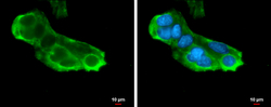
- RPL5 antibody detects RPL5 protein at Cytoplasm by immunofluorescent analysis. Sample: HepG2 cells were fixed in 4% paraformaldehyde at RT for 15 min.Green: RPL5 protein stained by RPL5 antibody (GTX101821) diluted at 1:500.Blue: Hoechst 33343 staining.

- Submitted by
- GeneTex (provider)
- Main image
- Experimental details
- RPL5 antibody detects RPL5 protein at cytoplasm by immunofluorescent analysis.Sample: HepG2 cells were fixed in 4% paraformaldehyde at RT for 15 min.Green: RPL5 protein stained by RPL5 antibody (GTX101821) diluted at 1:500.Blue: Hoechst 33342 staining.Scale bar = 10 £gm.

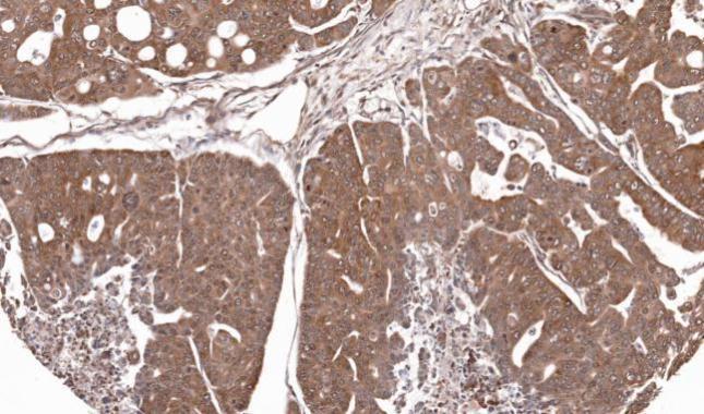
Supportive validation
- Submitted by
- GeneTex (provider)
- Main image
- Experimental details
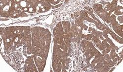
- Immunohistochemical analysis of paraffin-embedded Gastric CA N87 xenograft, using RPL5(GTX101821) antibody at 1:100 dilution.
- Submitted by
- GeneTex (provider)
- Main image
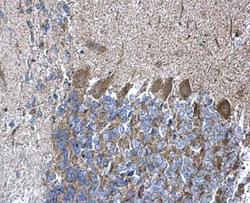
- Experimental details
- RPL5 antibody detects RPL5 protein at cytoplasm on mouse hind brain by immunohistochemical analysis. Sample: Paraffin-embedded mouse hind brain. RPL5 antibody (GTX101821) diluted at 1:500.
