Explore
 Validate
 Learn
Learn Western blot
Antibody data
- Antibody Data
- Antigen structure
- References [0]
- Comments [0]
- Validations
- Western blot [2]
- Immunocytochemistry [3]
- Immunohistochemistry [1]
Submit
Validation data
Reference
Comment
Report error
- Product number
- PA5-57857 - Provider product page

- Provider
- Invitrogen Antibodies
- Product name
- RIF1 Polyclonal Antibody
- Antibody type
- Polyclonal
- Antigen
- Recombinant full-length protein
- Description
- Immunogen sequence: ELNVGNEASF HGQERTKTGI SEEAAIEENK RNDDSEADTA KLNAKEVATE EFNSDISLSD NTTPVKLNAQ TEISEQTAAG ELDGGNDVSD LHSS Highest antigen sequence identity to the following orthologs: Mouse - 38%, Rat - 36%.
- Reactivity
- Human
- Host
- Rabbit
- Isotype
- IgG
- Vial size
- 100 µL
- Concentration
- 0.2 mg/mL
- Storage
- Store at 4°C short term. For long term storage, store at -20°C, avoiding freeze/thaw cycles.
No comments: Submit comment
Supportive validation
- Submitted by
- Invitrogen Antibodies (provider)
- Main image

- Experimental details
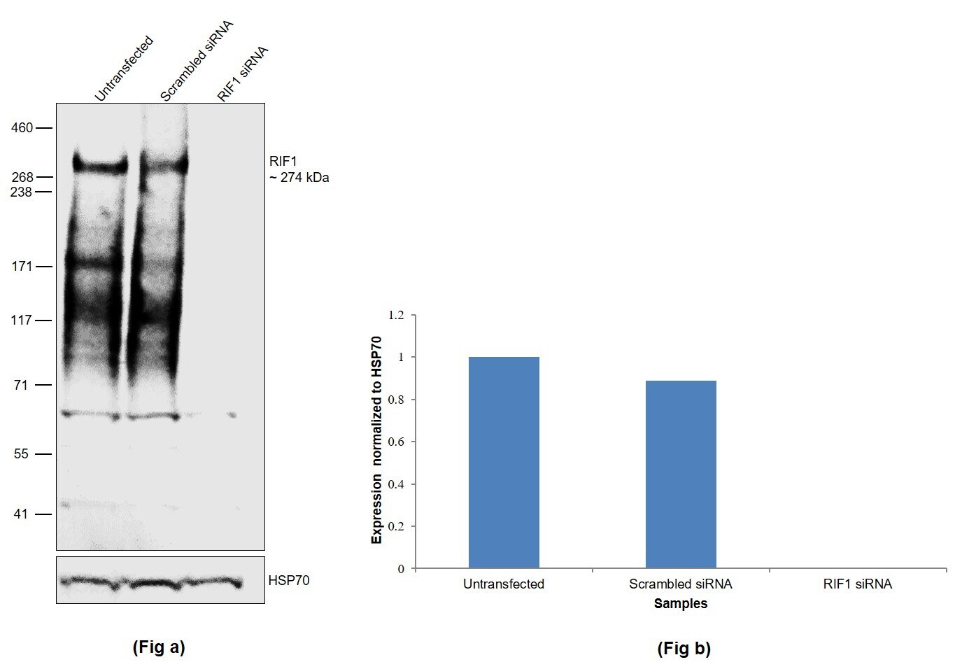
- Knockdown of Telomere-associated protein RIF1 was achieved by transfecting K-562 with Telomere-associated protein RIF1 specific siRNAs (Silencer® select Product # S30376, S30378). Western blot analysis (Fig. a) was performed using Nuclear enriched extracts from the Telomere-associated protein RIF1 knockdown cells (lane 3), non-targeting scrambled siRNA transfected cells (lane 2) and untransfected cells (lane 1). The blot was probed with RIF1 Polyclonal Antibody (Product # PA5-57857, 1:500 ) and Goat anti-Rabbit IgG (H+L) Superclonal™ Recombinant Secondary Antibody, HRP (Product # A27036, 1:4000). Densitometric analysis of this western blot is shown in histogram (Fig. b). Decrease in signal upon siRNA mediated knock down confirms that antibody is specific to Telomere-associated protein RIF1.
- Submitted by
- Invitrogen Antibodies (provider)
- Main image
- Experimental details
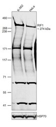
- Western blot was performed using Anti-RIF1 Polyclonal Antibody (Product # PA5-57857) and a 274 kDa band corresponding to Telomere-associated protein RIF1 was observed. Nuclear enriched extracts (30 µg lysate) of K-562 (Lane 1) and HeLa (Lane 2) were electrophoresed using NuPAGE™ 3-8% Tris-Acetate Protein Gel (Product # EA0378BOX). Resolved proteins were then transferred onto a Nitrocellulose membrane (Product # IB23001) by iBlot® 2 Dry Blotting System (Product # IB21001). The blot was probed with the primary antibody (1:500) and detected by chemiluminescence with Goat anti-Rabbit IgG (H+L) Superclonal™ Recombinant Secondary Antibody, HRP (Product # A27036, 1:4000) using the iBright FL 1000 (Product # A32752). Chemiluminescent detection was performed using Novex® ECL Chemiluminescent Substrate Reagent Kit (Product # WP20005). RIF1, being a telomere binding protein, interacts with a wide range of proteins which are involved in maintaining telomere stability and in controlling the replication process. Such interactions have high affinity and they are difficult to be dissociated under denaturing conditions. Hence, a RIF1 antibody detects multiple bands in Western Blot.
Supportive validation
- Submitted by
- Invitrogen Antibodies (provider)
- Main image

- Experimental details
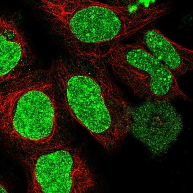
- Immunofluorescent staining of RIF1 in human cell line HEK 293 shows positivity in nucleus. Samples were probed using a RIF1 Polyclonal Antibody (Product # PA5-57857).
- Submitted by
- Invitrogen Antibodies (provider)
- Main image

- Experimental details
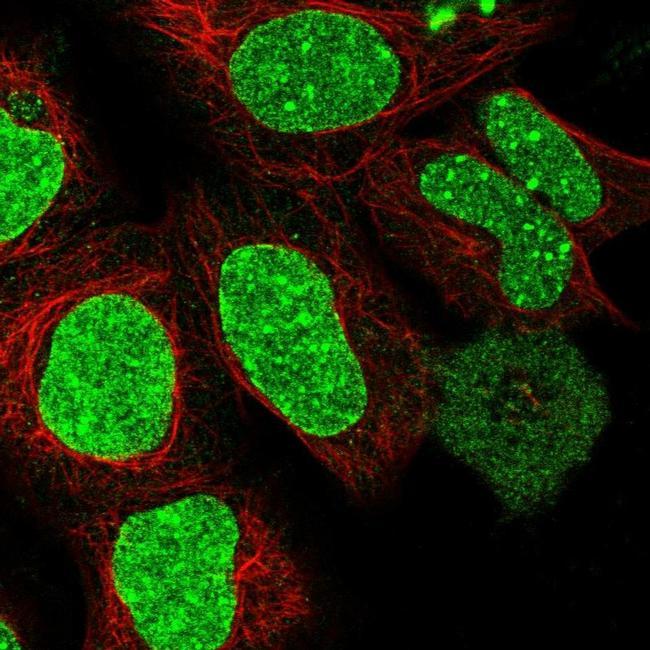
- Immunofluorescent staining of RIF1 in human cell line HEK 293 using a RIF1 Polyclonal Antibody (Product # PA5-57857) shows localization to nucleus, nuclear bodies and nuclear membrane.
- Submitted by
- Invitrogen Antibodies (provider)
- Main image

- Experimental details
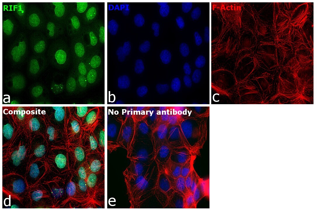
- Immunofluorescence analysis of RIF1 was performed using 70% confluent log phase NIH:OVCAR-3 cells. The cells were fixed with 4% paraformaldehyde for 10 minutes, permeabilized with 0.1% Triton™ X-100 for 15 minutes, and blocked with 2% BSA for 45 minutes at room temperature. The cells were labeled with RIF1 Polyclonal Antibody (Product # PA5-57857) at 1:200 in 0.1% BSA, incubated at 4 degree celsius overnight and then labeled with Donkey anti-Rabbit IgG (H+L) Highly Cross-Adsorbed Secondary Antibody, Alexa Fluor Plus 488 (Product # A32790), (1:2,000), for 45 minutes at room temperature (Panel a: Green). Nuclei (Panel b:Blue) were stained with ProLong™ Diamond Antifade Mountant with DAPI (Product # P36962). F-actin (Panel c: Red) was stained with Rhodamine Phalloidin (Product # R415, 1:300). Panel d represents the merged image showing nuclear localization. Panel e represents control cells with no primary antibody to assess background. The images were captured at 60X magnification in EVOS™ M7000 Imaging System (Product # AMF7000).
Supportive validation
- Submitted by
- Invitrogen Antibodies (provider)
- Main image
- Experimental details
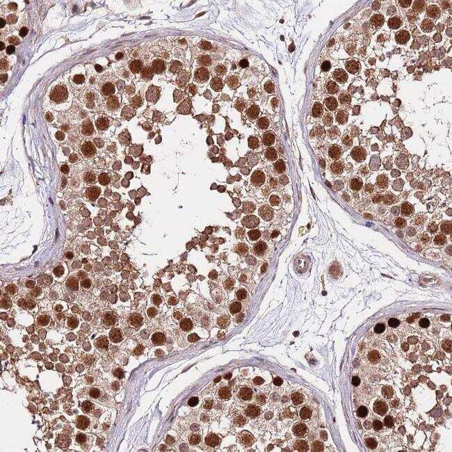
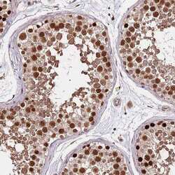
- Immunohistochemical staining of RIF1 in human testis tissue shows strong nuclear and cytoplasmic positivity in cells in seminiferus ducts. Samples were probed using a RIF1 Polyclonal Antibody (Product # PA5-57857).